How The Breast Biopsy Procedure Dictates Diagnostic Quality
Jun 14, 2026
https://my.clevelandclinic.org/health/diagnostics/24204-breast-biopsy-overview
The success of a breast biopsy procedure is ultimately judged by the pathologist examining the slide under the microscope. A well-designed procedure is the cornerstone of an accurate pathological diagnosis. Conversely, any flaw in the procedural execution can lead to a minute error resulting in a colossal mistake.
Step One: Quality Control in Specimen Acquisition
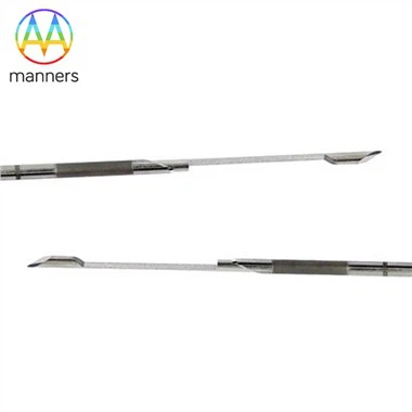
Pathologists dread receiving specimens that are "bloody messes" or "crushed fragments." Therefore, the puncture technique during the procedure directly impacts specimen quality. For instance, the core needle biopsy protocol demands hitting the target on the first pass-the needle tract must traverse the center of the lesion, avoiding areas of necrosis or hemorrhage. The ideal specimen is a complete, white tissue core, approximately 1.5–2 cm in length and 1–2 mm in diameter. While Vacuum-Assisted Biopsy (VAB) systems can harvest a large volume of tissue, excessive negative pressure or overly rapid rotation can cause crush artifact, distorting cellular morphology. The operator must adjust procedural parameters based on the hardness of the lesion (e.g., firmer in fibroadenomas, softer in invasive carcinomas).
Step Two: Standardized Specimen Handling Protocols
The procedure does not end once the specimen is removed. The tissue core must be immediately laid flat on filter paper saturated with formalin to prevent curling and desiccation. For microcalcifications, specimen radiography must be performed to confirm the presence of calcifications before sending the sample to pathology. Orientation marking is also crucial-using different colored dyes to mark the superior, inferior, medial, and lateral margins allows the pathologist to determine if the lesion was completely excised. This step is particularly vital in VAB procedures, where multiple specimens must be arranged in order to clarify the exact location of each tissue strip.
Step Three: The "Silent Dialogue" with Pathology
A high-quality pathology report relies heavily on the detailed information provided by the clinician on the requisition form: the imaging characteristics of the lesion (margins, shape, vascularity), the precise puncture site, and the clinical suspicion. For example, for a spiculated mass classified as BI-RADS 4C, the pathologist will focus on identifying features of invasive carcinoma; whereas for clustered fine calcifications, they will be highly vigilant for Ductal Carcinoma In Situ (DCIS). This transfer of information is an integral part of the procedure itself.
Step Four: New Specimen Requirements in the Molecular Era
With the advancement of precision medicine, the biopsy procedure must also reserve sufficient high-quality DNA/RNA for subsequent genetic testing (e.g., BRCA, PIK3CAmutations). This may necessitate increasing the number of passes (from the standard 3–4 to 5–6) and avoiding the use of instruments containing formaldehyde-based fixatives during the procedure to prevent nucleic acid degradation. The design of the procedure must be前瞻性 (forward-looking) to accommodate these potential needs.
In essence, the breast biopsy procedure is not merely a clinical task for the physician; it is a precision chain linking Radiology, Clinical Departments, and Pathology. The rigor applied at every step ultimately converges on a single goal: providing the patient with an indisputable pathological "gold standard" diagnosis.