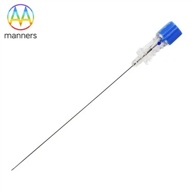
Histological Biopsy Needle PTC For Differential Diagnosis Of Cervical Inflammation And Precancerous Lesions
Nov 11, 2026
1. Clinical Pain Points
Cervical inflammatory erosion and precancerous lesions often look similar under 40× colposcopy, both showing red abnormal epithelial areas. CTC cytology can only find abnormal cells but cannot distinguish whether atypia comes from reactive inflammatory change or true dysplasia. Misdiagnosis brings two bad outcomes: treating benign inflammation as precancerous lesion leads to overtreatment, while missing high-grade CIN delays intervention before cancer develops. Non-diagnostic biopsy samples further prolong the diagnostic cycle. The challenge is to obtain layered tissue that shows epithelial architecture, nuclear atypia and stromal invasion, which cytology alone cannot provide. Inadequate or crushed biopsy specimens make this differential diagnosis impossible.
2. Working Principle
Histological biopsy needle PTC collects targeted tissue from colposcopy-suspicious cervical zones. After fixation and sectioning, pathologists observe epithelial stratification, nuclear morphology and mitotic activity under a microscope. Inflammatory lesions show reactive cell changes without disordered epithelial growth; precancerous lesions display progressive loss of polarity and atypical nuclear features. Biopsy is the only definitive diagnostic method when CTC is ambiguous. The stainless steel needle delivers precise, localized tissue capture. Smooth cutting edges preserve epithelial layers, so pathologists can judge whether the lesion is benign inflammation, low-grade dysplasia, high-grade dysplasia or invasive carcinoma. Colposcopy provides macroscopic guidance, while PTC biopsy provides histological evidence.
3. Classification of Histological Biopsy Needle PTC for Differential Diagnosis
Fine screening needles (0.25–1.2 mm): for borderline lesions with mild colposcopic abnormality and unclear CTC, low trauma, suitable for initial differentiation. Medium diagnostic needles (1.2–5 mm): for highly suspicious dysplasia, enough stromal tissue to evaluate lesion depth. Custom special needles: customized tip geometry for endocervical lesions invisible on the surface. Surface finish options: polishing, electroplating, plating and ultrasonic cleaning. ISO9001 certification, diameter range 0.25–30 mm, customizable by drawing or sample, packaging can be customized.
4. Operation Guide for Differential Diagnosis Biopsy
Preoperative review: CTC report, HPV status, colposcopic acetic acid and iodine staining findings. Mark the highest-risk area, usually the squamocolumnar transformation zone. Choose fine PTC needle for first sampling in borderline cases. Harvest one intact specimen from the most atypical region. Avoid sampling only obvious inflammatory areas while ignoring faint acetowhite spots. Keep the specimen intact and properly fixed. After pathology results return, classify lesions: benign inflammation, low-grade CIN, high-grade CIN or carcinoma. Adjust treatment plan accordingly: observation for inflammation, surveillance or excision for dysplasia. If the result conflicts with colposcopic impression, consider repeat biopsy with a customized needle.
5. Practical Experience
Reactive inflammatory atypia is the most common pitfall for CTC screening. Only histological sections can show that atypical cells are limited to inflamed surface layers without disordered proliferation. When the colposcopic image is discordant with cytology, the biopsy site selection is more important than taking more samples. Fine polished PTC needles are ideal for these differential cases because they produce intact epithelial layers without crush artifact. For patients with persistent positive CTC but mild colposcopy appearance, operators should avoid large needles that create unnecessary trauma. If the first biopsy only shows inflammation but clinical suspicion remains high, customized angled needles for endocervical sampling often identify hidden glandular lesions.
6. Summary
Histological biopsy needle PTC enables tissue-level differentiation between cervical inflammatory erosion and precancerous lesions. It resolves the limitation that CTC cytology cannot distinguish reactive atypia from true dysplasia. Graded needle specifications match borderline screening and high-suspicion scenarios. Intact specimens captured by PTC needles provide reliable evidence for pathology grading, preventing both overtreatment and missed diagnosis.
7. Prospect and Suggestion
Future PTC biopsy needles can combine micro-sampling design for multi-site minimally invasive collection. Manufacturers should maintain stable sharpness for fine differential-diagnosis needles. Gynecology departments should build image and pathology databases for inflammatory and dysplastic lesions for clinician training. For patients with persistent atypical cytology and inconclusive colposcopy, PTC biopsy should be prioritized. Medical device suppliers need to provide fast customized service for endocervical sampling to reduce false-negative rates.
< img alt="news-1-1" src="/uploads/24240/news/202604241539186d0ff.png " />