Franseen Crown And Crown Core Cut Designs From A Crown End Needles Manufacturer’s Perspective
Jul 23, 2026
Acquiring high-quality tissue specimens is paramount for definitive diagnosis and treatment planning in solid tumor and deep-organ biopsies. Conventional bevel-tipped needles often encounter challenges such as tissue slippage, specimen fragmentation, or insufficient sampling-particularly within fibrotic or dense lesions. Addressing these limitations, novel biopsy needle geometries like the Franseen (Crown) and Crown Core Cut designs have emerged. Their defining feature-multi-pronged grasping structures-significantly enhances "bite stability," securing larger, intact tissue cores. As a Crown End Needles Manufacturer specializing in high-end devices, fabricating these complex geometries presents unprecedented precision machining challenges. It demands not only micron-level machining capabilities but also a profound understanding of material behavior, force transmission, and tissue-needle interactions.
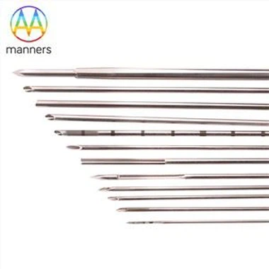
The Franseen Needle, named for its resemblance to a crown with three (or more) radiating cutting prongs, draws inspiration from natural predatory tools. Unlike traditional bevels or cones, the Franseen tip comprises multiple sharp cutting teeth separated by intervening valleys. Typically spaced 120 degrees apart, each tooth features a keen leading edge and a supportive trailing edge. During rotational or forward advancement, these teeth act as synchronized micro-blades. Crucially, this multi-tooth configuration generates powerful "bite" or "grip" forces. Upon tissue contact, the three teeth engage the lesion from multiple vectors, forming a stable triangular purchase that prevents the needle from skidding or drifting within fibrous tissue. This stability allows precise trajectory control. Once suction or cutting commences, the engaged tissue is securely held within the "crown," enabling the needle to excise a clean, cylindrical core as it advances. From a manufacturing viewpoint, the primary challenge lies in achieving perfect symmetry and uniform sharpness across all prongs. Identical tooth length, angle, and curvature radius are essential for balanced force distribution and straight tracking. Minute discrepancies can cause uneven loading, tip deflection, or prong fracture. We employ advanced micro-Electrical Discharge Machining (micro-EDM) or femtosecond laser cutting to form the intricate Franseen crown structure. These non-contact methods prevent mechanical stress and heat-affected zones while enabling precise geometry replication. Subsequent multi-stage electropolishing removes micro-burrs from the prong tips, achieving mirror-like sharpness while ensuring smooth, radiused transitions at the prong roots to prevent stress concentrations.
Crown Core Cut needles represent another innovation targeting improved tissue yield. Sharing conceptual similarities with Franseen needles (multi-lobe tips), they differ in prong configuration and cutting mechanics. The Crown Core Cut tip may feature two, three, or four petal-like lobes. In the closed position, these lobes form a continuous cone or pyramid for tissue penetration. During tissue acquisition (via cutting or aspiration), the lobes may flare open or remain fixed, presenting a larger cutting circumference. The core advantage lies in its "embracing" capacity. Once within the target, the lobed structure envelops the tissue. Rotational or axial motion then excises a core encircled by the petals. Compared to single-bevel needles, the Crown Core Cut offers a larger cutting surface area and superior tissue retention. This proves particularly advantageous for fragile or friable lesions (e.g., lymphoma), significantly increasing the likelihood of obtaining intact, diagnostically useful cores suitable for molecular pathology. Manufacturing Crown Core Cut needles centers on precision lobe forming and, for deployable versions, reliable actuation mechanisms. Fixed-crown variants require exacting control over lobe-tip sharpness and inter-lobe spacing-too narrow impedes tissue entry, too wide risks specimen fragmentation. Precision micro-grinding with custom form tools shapes the complex lobe contours. Deployable Crown Core Cut needles add complexity: internal push-rods, linkages, and micro-assembly are needed to ensure smooth actuation, precise positioning, and consistent repeatability.
Clinically, Franseen and Crown Core Cut needles dominate Endoscopic Ultrasound-guided Fine Needle Biopsy (EUS-FNB), percutaneous liver biopsy, and pancreatic mass sampling. These scenarios involve deep-seated targets surrounded by critical anatomy, making first-pass success rates vital. Franseen needles excel in fibrotic lesions like pancreatic cancer, where their bite stability overcomes tissue hardness and prevents slippage. Crown Core Cut needles demonstrate superiority in softer tissues (e.g., lymphoma), where their larger cutting diameter and tissue-retentive geometry capture longer, more architecturally preserved cores. As precision medicine advances, demanding not just quantity but also structural integrity and cellular viability for molecular analyses, the clinical need for these high-performance biopsy needles intensifies.
Quality assurance for these complex needles is paramount. We operate dedicated micro-inspection labs equipped with Scanning Electron Microscopes (SEM) and white light interferometers. For Franseen needles, we rigorously inspect prong symmetry, tip radius, edge quality, and surface roughness. Simulated biopsy testing using biomimetic tissue analogs assesses bite efficacy and sample quality. For Crown Core Cut needles, besides geometric verification, we subject deployment mechanisms to thousands of durability cycles to preclude intraprocedural malfunction. All products undergo stringent passivation and final sterile packaging under our ISO 13485-compliant Quality Management System (QMS), ensuring traceability from raw material to finished device.
Future trends in tissue biopsy point toward greater precision, safety, and minimally invasiveness. Confocal laser endomicroscopy integrated with biopsy needles may enable "optical biopsy" concurrent with tissue acquisition. Magnetic navigation promises enhanced targeting accuracy. As a responsive Crown End Needles Manufacturer, we align our R&D with these trajectories. We investigate novel alloys and surface enhancements to boost tip hardness and wear resistance. We explore AI-driven automated optical inspection (AOI) for micron-level defect detection. Our overarching goal is to empower clinicians with increasingly sophisticated biopsy tools. Each precise bite we enable supports accurate diagnosis and effective treatment. We believe that through relentless innovation and uncompromising quality, we play a vital role in advancing patient care.