Ensuring The Accuracy Of Breast Biopsy Results Through Full-Chain Quality Control
Jun 27, 2026
https://www.sirius-medical.com/knowledge/breast-biopsy-needle-techniques
Keywords: Breast biopsy results, accuracy, quality control
The accuracy of breast biopsy results is a lifeline concerning patients' lives and health. A false-negative result may lead to delayed treatment, while a false-positive result may trigger unnecessary surgery and psychological trauma. Therefore, from the selection of biopsy needles and puncture operation to specimen processing and pathological diagnosis, every link in the entire chain must strive for excellence to ensure that the final breast biopsy result issued is authentic and reliable.
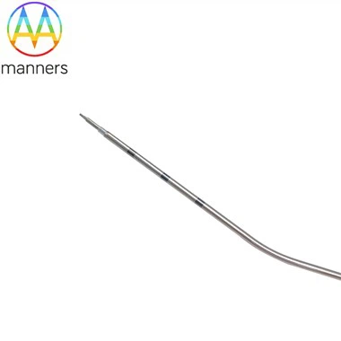
First, the sampling phase is the starting point of quality. As mentioned earlier, the material of the biopsy needle (stainless steel, titanium alloy, medical plastic) and specifications (length, diameter/gauge) directly affect the quality of sampling. For example, for microcalcifications, a vacuum-assisted biopsy system may be needed to obtain larger, more complete tissue strips; whereas for cystic lesions, fine-needle aspiration may suffice. The experience and skill of the physician are also crucial-whether the biopsy needle can be precisely placed at the center of the target lesion under image guidance directly determines the representativeness of the sampled tissue. The extracted tissue should be properly fixed immediately to prevent autolysis or degradation.
Second, the specimen processing and slide preparation stages are equally critical. Pathological technicians need to perform a series of complex procedures on the extracted tiny tissue blocks, including dehydration, embedding, sectioning, and staining. Any mistake in any step, such as uneven section thickness or over-/under-staining, may affect the pathologist's interpretation. Especially for specimens requiring HER2 FISH testing, good tissue fixation and section quality are prerequisites for success.
Third, pathological diagnosis is the core link. Pathologists need to carefully observe cellular morphology, structural arrangement, nuclear atypia, and other characteristics under a microscope to make a judgment on benignity or malignancy. For difficult cases, immunohistochemical staining may be needed to assist in diagnosis. For example, markers such as CK5/6 and E-cadherin are used to differentiate ductal carcinoma in situ from lobular carcinoma in situ. An accurate pathological report should not only have a clear diagnostic conclusion, but also include all necessary grading, staging, and molecular marker information.
Fourth, quality control and multidisciplinary discussion are the final safeguards. Regular pathology departments establish strict internal quality control systems, including periodic reviews and consultations on difficult cases. More importantly, breast biopsy results should not be viewed in isolation. They must be combined with the patient's clinical presentation and imaging characteristics (such as mammography, ultrasound, MRI) for multidisciplinary comprehensive discussion. For example, if a lesion that is highly suspicious for malignancy on imaging yields a benign biopsy result, it is necessary to re-evaluate: Was the sampling inadequate? Is a repeat biopsy or surgical excisional biopsy needed? This principle of "clinical-imaging-pathology" correlation is the last line of defense against misdiagnosis and missed diagnosis.
In short, a reliable breast biopsy result is the product of the combined effect of technology, experience, and strict management. It embodies the professional collaboration of multiple teams including radiologists, surgeons, pathology technicians, and pathologists. Only by establishing a full-chain quality control system from sampling to interpretation can we ensure that every breast biopsy result stands up to scrutiny and becomes a solid basis for guiding clinical decision-making.