Cytological Pitfalls And Accuracy Control Systems in FNAB
Jul 18, 2026
https://www.mayoclinic.org/tests-procedures/breast-biopsy/about/pac-20384812
The diagnostic foundation of Fine Needle Aspiration Biopsy (FNAB) rests upon cytological slide interpretation. This diverges fundamentally from the histopathological analysis of core biopsies, relying solely on dispersed free cells or cell clusters without the contextual clues of intact tissue architecture. Consequently, FNAB harbors unique diagnostic pitfalls and quality control (QC) challenges. Establishing standardized accuracy control systems to navigate these cytological complexities is paramount to safeguarding the clinical utility of FNAB and minimizing misdiagnosis or missed diagnoses.
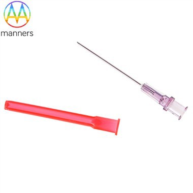
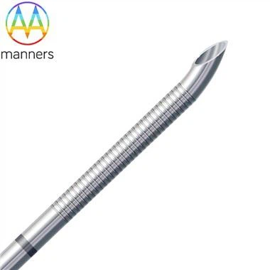
Sampling error represents the most frequent primary QC issue and the leading cause of false-negative results. FNAB, utilizing an ultra-fine needle, retrieves only locally available free cells. If the needle trajectory deviates, fails to penetrate the tumor core, or solely aspirates surrounding normal adipose or fibrous stroma, the specimen becomes non-diagnostic. This risk escalates with minute lesions, densely fibrotic masses, or clusters of microcalcifications, where the fine needle struggles to capture diagnostic atypical cells, potentially leading to under-detection of malignancy. Furthermore, suboptimal intraoperative negative pressure regulation can result in blood dilution of the sample, depleting the concentration of diagnostically relevant cells-a prevalent QC shortfall in primary care settings.
The specialized expertise demanded for cytological interpretation and its inherent diagnostic traps constitute a core QC challenge. Unlike histology, which permits assessment of basement membranes, glandular structures, and tissue organization, FNAB cytology hinges entirely on evaluating individual cell characteristics: size, nuclear-to-cytoplasmic ratio, chromatin texture, and degree of atypia. This imposes rigorous demands on pathologist experience. Diagnosing Ductal Carcinoma In Situ (DCIS) epitomizes this difficulty; confined within the ductal lumen, DCIS cells often exhibit subtle atypia without architectural context, rendering them misclassified as benign hyperplasia or usual ductal hyperplasia. Conversely, certain benign atypical hyperplasias may display sufficient cytological atypia to be over-called as malignant, precipitating overtreatment. Papillary lesions and inflammatory processes further complicate interpretation due to overlapping cytological features and blurred diagnostic boundaries.
Non-adherence to standardized sample handling protocols amplifies diagnostic errors. The liquid-based cytology specimen obtained via FNAB is highly perishable, demanding stringent timeliness and preparation techniques. Delayed fixation, uneven smear thickness, or inconsistent staining artifacts degrade cellular morphology, obfuscating interpretation. Overly thick smears cause cellular crowding and obscuration, hindering single-cell appraisal. Failure to fix specimens promptly invites air-drying artifact or autolysis, destroying nuclear detail and directly undermining diagnostic accuracy-entirely preventable pre-analytical errors.
A robust QC infrastructure is the cornerstone for elevating diagnostic precision. Standardized clinical protocols mandate strict adherence to multi-pass fanning sampling to ensure specimen representativeness. Promoting ROSE allows immediate verification of sample adequacy at the point of care, enabling instantaneous remedial passes if indicated-addressing sampling errors at their source. Establishing dedicated cytopathology training programs focused on refining criteria for diagnosing DCIS, atypical hyperplasia, and inflammatory mimics is essential. Furthermore, adopting a multidisciplinary approach by correlating cytological findings with BI-RADS imaging categories is crucial; imaging-highly-suspicious lesions with discordant negative cytology warrant escalation to core needle biopsy. Implementing this closed-loop QC system can stabilize FNAB diagnostic accuracy above 90%, effectively mitigating clinical diagnostic risks.