CT And Ultrasound Dual-Guided Positioning Performance
Oct 01, 2026
1. Industry Dual-Guided Positioning Pain Points
Traditional brachytherapy particle implantation instruments have poor adaptive matching with CT and B-ultrasound dual guidance modes, resulting in prominent positioning accuracy defects. Ordinary puncture needles have uneven structural density and rough surface, easy to generate strong artifacts and echo interference under ultrasound imaging, blurring lesion boundaries and needle tip positions. Under CT scanning, traditional needles have inconsistent imaging signals, prone to position deviation judgment, unable to achieve millimeter-level precise positioning. Most traditional products have no unified dimensional calibration standard, and the needle body depth identification is fuzzy, making it difficult to accurately control puncture depth under dual-mode guidance. In addition, traditional instruments cannot switch adaptively between CT and ultrasound guidance modes, with single-scene applicability. Blind puncture and positioning deviation easily lead to particle implantation offset, uneven radiotherapy dose distribution, missed treatment of tiny lesions and excessive radiation damage to normal tissues, seriously affecting the therapeutic effect of precise brachytherapy.
2. Core Dual-Guided Positioning Working Principle
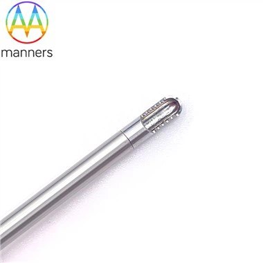
Oncology Brachytherapy Particle Implant Needles are professionally optimized for CT and B-ultrasound dual-mode imaging guidance, with excellent multi-scene positioning adaptability. The high-precision integrated forming technology of necking and swaging ensures uniform needle body density and stable structural consistency, enabling linear and uniform propagation of ultrasonic signals under ultrasound guidance, reducing echo artifacts and interference, and realizing clear needle tip and lesion imaging. The precise laser marking process forms high-resolution depth scales on the needle body, matching CT three-dimensional scanning data and ultrasound real-time dynamic positioning data, realizing accurate closed-loop control of puncture depth and angle. The electropolished smooth surface eliminates surface micro-protrusions that cause imaging interference, ensuring stable and consistent imaging effects under dual guidance modes. The full-specification graded design adapts to different tissue imaging characteristics, realizing seamless switching and precise positioning of CT stereotactic scanning and ultrasound real-time monitoring.
3. Dual-Guided Adaptive Product Classification
According to dual-mode imaging adaptability and positioning precision, the products are divided into ultrasound real-time monitoring type and CT stereotactic high-precision type. The ultrasound real-time monitoring type optimizes surface smoothness and structural uniformity, with weak echo interference and high real-time imaging clarity, suitable for superficial tumor dynamic puncture and real-time particle implantation adjustment. The CT stereotactic high-precision type adopts ultra-precision dimensional calibration and anti-artifact structural design, with high positioning repeatability and millimeter-level precision, specially suitable for deep visceral tumor three-dimensional stereotactic implantation and tiny lesion precise radiotherapy. All products support customized structural optimization for dual-mode imaging, covering all clinical conventional and high-precision implantation scenarios.
4. Dual-Guided Standard Operation Guidelines
Ultrasound-guided operation: Select ultrasound-adaptive implant needles, adjust probe frequency and gain parameters to clarify tumor lesion, peripheral blood vessels and needle body imaging. Puncture slowly along the ultrasound long-axis perspective path, dynamically observe needle tip advancement trajectory, adjust angle in real time to avoid vascular injury. Stabilize the needle body after reaching the target lesion to complete uniform particle implantation. CT-guided operation: Preoperatively perform multi-layer CT scanning to delineate tumor target area, formulate three-dimensional puncture depth and angle scheme. Calibrate needle body scale and CT stereotactic data to ensure data consistency. Adopt low-speed stable puncture according to preoperative plan, verify needle tip position through real-time CT scanning, and complete fixed-point quantitative particle implantation to ensure accurate dose distribution.
5. Practical Dual-Guided Application Experience
Clinical dual-mode guided brachytherapy verification proves that Oncology Brachytherapy Particle Implant Needles have outstanding positioning stability and scenario adaptability. In ultrasound dynamic implantation, the product has negligible artifact interference, clear and intuitive needle tip imaging, effectively avoiding positioning deviation caused by blurred vision, and improving the real-time adjustment efficiency of particle distribution. In CT stereotactic precise implantation, the precise scale calibration and uniform structural density perfectly match three-dimensional positioning data, realizing accurate implantation of millimeter-level tiny deep lesions that are difficult to locate with traditional needles. The dual-mode adaptive design enables free switching of guidance modes according to lesion location and surgical needs, greatly expanding clinical application scenarios. The accurate positioning performance effectively optimizes radiotherapy dose conformity, improves tumor local control rate, and reduces normal tissue radiation damage, obtaining unanimous recognition from clinical oncology and imaging departments.
6. Summary and Sublimation
Oncology Brachytherapy Particle Implant Needles effectively solve the industry pain points of serious imaging artifact interference, poor dual-mode adaptability and low positioning accuracy of traditional brachytherapy instruments. Through structural uniformity optimization, precision scale calibration and anti-artifact design, it realizes perfect matching with CT and ultrasound dual guidance systems. It improves the precision, stability and scenario flexibility of tumor particle implantation surgery, avoids treatment errors and dose deviation caused by positioning inaccuracy, provides reliable instrument support for dual-mode guided precise brachytherapy, and promotes the standardized and high-precision development of modern tumor radiotherapy technology.
7. Industry Dual-Guided Technology Development Suggestions
The industry should continue to optimize the dual-mode imaging adaptive design of brachytherapy needles, develop low-artifact structural processing technology suitable for complex tissue imaging. Enterprises should enrich dual-mode adaptive product lines, strengthen customized optimization for deep complex lesions and tiny micro-lesion implantation scenarios. Medical institutions should formulate unified dual-guided brachytherapy operation specifications, standardize the matching scheme of imaging equipment and needle specifications. Strengthen multi-center dual-mode contrast clinical research, continuously improve the positioning precision and scenario adaptability of domestic implant needles, and lead the technical upgrading of precise brachytherapy industry.