Clinical Operating Specifications And Precise Puncture Techniques For AVF Cannulation
Jun 04, 2026
https://www.kidney.org/sites/default/files/Fistula%20Bulletin.pdf
Arteriovenous Fistula (AVF) serves as the "lifeline" for patients receiving maintenance hemodialysis. Successful AVF cannulation marks the critical initial step to guarantee adequate dialysis efficacy and prolong fistula service life. Far from a routine puncture procedure, AVF cannulation is an intricate clinical practice integrating anatomical expertise, refined operative proficiency and patient-centered humanistic care. This article elaborates on core clinical specifications and key precise puncture techniques for AVF cannulation.
I. Pre-cannulation Comprehensive Assessment and Planning: Foundation for Successful Cannulation
Successful cannulation hinges on thorough preoperative preparation, starting with maturation evaluation of the target AVF. A mature fistula presents prominent thrills, distinct vascular bruits, sufficient blood flow (typically above 600 mL/min), and superficially positioned, arterialized vascular walls. Through inspection, palpation, auscultation combined with ultrasonic examination, clinicians can accurately measure fistula diameter, subcutaneous depth, vascular course and the usable length of puncture segments. Ultrasound, regarded as the gold standard for evaluation, intuitively visualizes inner lumen diameter, intimal integrity, and the presence of vascular stenosis or thrombosis.
Next comes puncture site planning. Cannulation shall follow the rope-ladder or buttonhole puncture protocols to avoid aneurysmal dilatation or vascular stenosis induced by localized repetitive puncturing. The primary puncture site shall be positioned a minimum of 3 cm away from the anastomosis on straight, elastic vascular segments. The arterial needle (blood outflow end) is generally directed proximally, while the venous needle (blood return end) points distally; the inter-needle spacing is recommended to exceed 5 cm to mitigate blood recirculation rate.
II. Core Technical Procedures for Precise Puncture
1. Selection of Puncture Needles
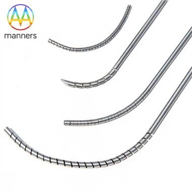
As specified in product documentation, specialized AVF cannulation needles (e.g., 17G specification with an outer diameter of 1.45 mm and inner diameter of 1.19 mm) form the material prerequisite for standardized puncture. Fabricated from 316L stainless steel, these needles deliver superior biocompatibility and corrosion resistance under in-vivo blood environment. Tip grinding is a pivotal manufacturing process: finished tips feature ultra-high sharpness (penetration force ranging from 50 g to 100 g) and smooth finish to minimize intimal trauma and puncture-associated pain. Advanced five-axis laser cutting is adopted to fabricate lateral openings, optimizing blood flow and reducing the risk of needle apposition against vessel wall, a hallmark of premium-grade cannulation needles.
2. Aseptic Manipulation and Skin Disinfection
Rigorous aseptic technique is essential to prevent infectious complications including bacteremia and subcutaneous abscesses. Full standardized disinfection is implemented over the puncture field followed by sterile drape placement; operators must don sterile gloves prior to manipulation.
3. Puncture Angle and Needle Insertion Maneuvers
Conventional puncture angle ranges from 20° to 30°. For superficially located fistulas, a reduced insertion angle of 15°–20° or a tangential nicking technique can be applied to lower the risk of posterior vessel wall perforation. During advancement, the needle bevel is kept upward; once the characteristic "give" sensation of lumen penetration is felt, the needle hub is flattened and advanced parallel along the vascular course for a short distance. Ultrasound-Guided Puncture (UGP) has evolved into gold-standard practice, enabling real-time visualization of needle tip positioning to achieve single-pass intraluminal cannulation. Particularly indicated for newly matured fistulas, deep-set fistulas or patients with difficult vascular access, UGP drastically improves puncture success rate and curbs complications such as hematoma.
4. Needle Securement and Tubing Connection
Upon successful cannulation, dedicated adhesive dressings are used to fix needle wings firmly against skin to prevent needle displacement or dislodgement. Strict air purging is required when connecting extracorporeal blood circuits, with tight junctions secured to avoid air embolism.
III. Post-cannulation Immediate Assessment and Monitoring
Immediate post-procedural evaluation is mandatory after cannulation completion, covering patency, color and pressure of returning blood alongside unchanged auscultated vascular bruits. After connection to the hemodialysis machine, continuous monitoring of arterial pressure, venous pressure and actual blood flow is performed to verify compliance with preset targets. Abnormal readouts often signal malpositioned needle, vessel-wall apposition or intrinsic vascular lesions.
Conclusion
Precise AVF cannulation constitutes a systematic workflow encompassing preoperative assessment, site planning, operative proficiency and appropriate instrumentation. Practitioners are required to command solid theoretical foundations and polished hands-on skills, alongside rigorous professionalism and robust accountability for patient safety. Driven by the popularization of visualized modalities such as ultrasound guidance and the clinical adoption of high-performance cannulation needles manufactured via precision laser machining, AVF puncture continues to evolve toward enhanced accuracy, safety and comfort. The ultimate objective is to maximally preserve patients' vascular lifelines, improve dialysis outcomes and elevate overall quality of life.