Chiba Needle Tip Visualization Under Image Guidance And Puncture Path Control
Jul 04, 2026
Needle Tip Identification and Deflection Correction Under Ultrasound/CT/MRI Guidance
https://radiopaedia.org/articles/chiba-needle
The most widespread application of the Chiba needle is percutaneous biopsy, cyst aspiration, PTCD, and nerve blocks under image guidance. In these scenarios, the tip's visibility under imaging (tip visibility) and the deflection behavior caused by the bevel are knowledge that operators must master.
Needle Tip Display Under Ultrasound Guidance
High-frequency linear array probes (5–12 MHz) are commonly used for superficial organ biopsies (thyroid, breast, lymph nodes). Standard Chiba needles are metallic and appear as strong hyperechoic bright lines on ultrasound, with the tip showing a "comet-tail" artifact or bright dot. However, because 21G–23G Chiba needles are very thin (<0.9 mm), the needle body tends to "disappear" when the sound beam angle relative to the needle is too large. Techniques to enhance tip visibility include:
Adjusting the incident angle so the sound beam is approximately parallel to the needle body (in-plane technique);
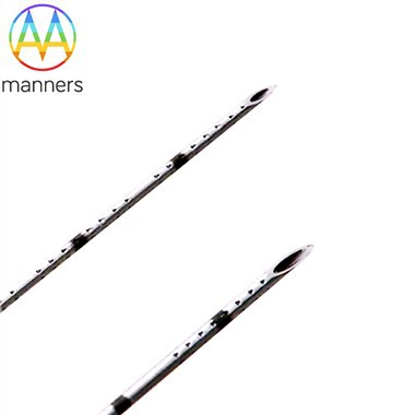
Selecting Chiba needles with echogenic tip coating or micro-grooved tips, where laser-etched micro-grooves at the tip create more scattered echoes;
Gently moving the needle tip in and out to confirm position.
Needle Tip Localization Under CT/MRI Guidance
CT guidance is mostly used for deep lung, liver, and retroperitoneal lesions. The Chiba needle contains stainless steel, appearing as a very high-density dot on CT, easy to track. The "stepwise advancement - scan confirmation - further advancement" strategy is typically used, checking with a CT slice every 1–2 cm of advancement to ensure the planned path is not deviated.
In MRI environments, non-ferromagnetic Chiba needles (eitanium alloy or specific stainless steel, labeled MRI Compatible) must be used to avoid magnetic field disturbance and image artifacts. Some改良 Chiba stylets contain paramagnetic markers to assist MR fluoroscopic navigation.
Bevel Deflection Effect and Clinical Countermeasures
When the Chiba needle bevel faces a certain direction, the needle tip will bend slightly toward the opening side during puncture (typically 0.5°–2°/cm, depending on tissue density and insertion speed). Operational key points:
When the planned path is close to important vessels/airways, actively set the bevel to face the safe side (bevel-away from critical structures);
To correct deflection, rotate the needle body 180° to reverse the deflection direction, or withdraw slightly and reorient;
For deep tiny lesions (<1 cm), the coaxial technique is recommended - first place the Chiba needle near the lesion, then exchange for a larger cutting needle for sampling, reducing needle drift caused by repeated punctures.
"Give-Way Sensation" and Penetration Confirmation
When the Chiba needle punctures into a fluid-filled lesion (cyst, abscess) or enters a low-pressure bile duct, the operator often feels a slight "give-way sensation." Aspiration with a syringe to retrieve contents confirms the tip is in place. In PTCD procedures, a small amount of contrast is injected to confirm the tip is within the biliary tree before guidewire insertion.
Mastering the performance characteristics of the Chiba needle tip under different imaging modalities and the规律 of bevel deflection can significantly improve first-attempt puncture success rates and reduce complications such as pneumothorax and bleeding.