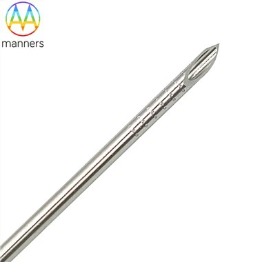
Bevel Geometry And Tip Mechanics
Nov 11, 2026
The disposable PTC aspiration biopsy needle looks deceptively simple, yet its clinical behavior is governed almost entirely by the geometry of its tip. In interventional radiology, the difference between a clean first-pass duct entry and a wandering, traumatic puncture often comes down to a few degrees of bevel angle and a few microns of edge finish. The pain point is consistency: operators expect every needle from every box to feel identical, but many procurement teams treat needles as commodities and ignore the manufacturing tolerance that defines tip performance.
From the perspective of structural mechanics, the bevel is not merely a sharpened end-it is a load-directing wedge. A standard Chiba-style PTC needle uses a long bevel ground at 25° to 30°. This angle converts the axial force from the operator's hand into a controlled piercing moment. A smaller bevel angle penetrates deeper with less lateral dissection, which is useful in dense hepatic fibrosis, but it can feel "tip-heavy" and drift off-axis. A larger bevel separates tissue more gently and gives better tactile feedback, but increases the effective contact area and may require more push in scarred tissue. The ideal compromise for biliary access is a 25° to 28° machine-cut bevel with a symmetric heel and no burr.
Material selection is the first layer of performance. Most PTC needles use 304 or 316L stainless steel thin-wall tubing. 316L offers better corrosion resistance and lower extractables, which matters for cytology where metal ions could affect staining. Nitinol shafts are used in premium lines for kink resistance and MRI compatibility, but Nitinol is harder to grind consistently and requires strict nickel-surface control. The tube must be drawn to tight concentricity: if the wall thickness varies around the circumference, the bevel will cut unevenly and the tip will deflect toward the thinner side.
The stylet is an overlooked component. In a quality PTC set, the stylet fits the lumen with a clearance small enough to block tissue ingress but loose enough to withdraw smoothly. During capsular crossing, the stylet prevents plugging and stabilizes the tip. Once withdrawn, the hollow cannula becomes the aspiration channel. If the stylet tip is recessed or proud relative to the cannula bevel, the puncture feel changes and the risk of capsular skipping rises.
Surface finish determines friction. An electropolished inner lumen reduces dead space and prevents cell clinging, which improves sample recovery in thyroid, lymph node, and pancreatic FNA. Outer-surface polishing lowers tissue drag, but a fully mirror-polished shaft becomes invisible under ultrasound. That is why echogenic treatment is concentrated near the tip: micro-etching, laser dimples, or helical roughening create controlled acoustic scatter without compromising penetration.
Device classification by tip design includes the standard 25° Chiba bevel for general PTC and FNA; the ultra-fine 22G to 25G bevel for superficial cytology; the echo-ring tip with spiral microtexture for real-time ultrasound guidance; and the flush-aligned coaxial bevel where inner needle and outer sheath edges meet precisely to avoid a step that could catch on tissue. Each variant is a trade-off between sharpness, visibility, and trackability.
In practice, tip inspection should be part of incoming QC, not just final packaging. Optical profilometry can measure bevel angle, edge radius, and symmetric land width. A good tip has a continuous cutting edge with radius in the low-micron range and no rolled heel. Operators can sense this immediately: a quality tip enters with a clean "give" at the capsule; a dull or asymmetric tip judders, walks laterally, and forces correction passes.
The operation logic is straightforward. Confirm bevel orientation relative to the imaging plane, keep the stylet seated until the liver capsule is reached, advance with short steady pressure rather than a single hard thrust, and avoid rocking once the tip is in the target. Rocking a poorly finished bevel enlarges the track and raises bleeding risk.
Field experience shows that tip consistency is a training accelerator. When needles behave predictably, junior operators learn trajectory control faster because the variable of "bad tip" is removed. Conversely, intermittent quality erodes trust in the whole procedure. Departments that audit needle tip quality report fewer redirections and lower contrast volumes.
In summary, the bevel is the DNA of the PTC needle. It defines penetration vector, tactile feedback, sample integrity, and safety margin. Manufacturers should treat tip grinding as a precision process with statistical control, not a secondary finishing step.
Looking forward, the industry should move toward laser-defined bevels with closed-loop optical inspection, documented angle tolerance per batch, and patient-specific tip libraries-for example, finer bevels for pediatric or cirrhotic livers and slightly longer bevels for post-surgical fibrosis. The needle that feels invisible in the hand is actually the product of very visible engineering discipline.
< img alt="news-1-1" src="/uploads/24240/news/202604241539186d0ff.png " />