A Technological Leap From Blind Insertion To Precise Positioning
Jul 16, 2026
https://www.mayoclinic.org/tests-procedures/breast-biopsy/about/pac-20384812
The development of breast biopsy needle types has always been closely linked to image-guided technology. From the early "blind insertion" to today's real-time guidance from ultrasound, mammography, and MRI, the design iterations of biopsy needles have always revolved around the core goal of "precise positioning."
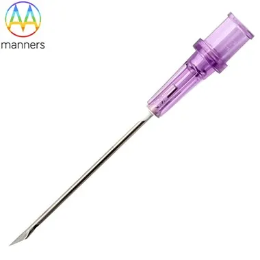
Ultrasound-guided biopsy needles are currently the most widely used type. Ultrasound can clearly display the size, shape, and blood flow signals of lesions, guiding doctors to accurately insert the biopsy needle into the target area. For this scenario, specialized biopsy needles typically feature an "acoustic enhancement design"-the needle surface is specially polished or coated with a metal layer, producing a strong echo in ultrasound images, facilitating real-time tracking of the needle tip position. For example, a certain brand's 14G ultrasound-guided biopsy needle uses a spiral tip design, which enhances ultrasound imaging while reducing tissue resistance during puncture. Furthermore, for patients with dense breast tissue, a slender (15cm) ultrasound-guided needle can penetrate deep tissue, avoiding damage caused by multiple punctures.
Mammography stereotactic biopsy needles are specifically designed for microcalcifications. Mammography can detect clusters of calcifications smaller than 5mm, but it cannot directly visualize the needle path; therefore, the biopsy needle needs a "positioning memory function." These needles typically integrate markers (such as small metal rings) at the needle hub, which, in conjunction with the stereotactic positioning system of the mammography machine, can accurately calculate the needle insertion angle and depth. For example, the application of vacuum-assisted biopsy (VAB) systems under mammography guidance, through continuous negative pressure suction, can acquire multiple tissue samples at once, achieving a calcification detection rate of up to 92%, significantly superior to traditional hollow needles.
MRI-compatible biopsy needles have become a technological hotspot in recent years. MRI is radiation-free and offers high soft tissue resolution, but the strong magnetic field environment places stringent requirements on the biopsy needle material-it must be completely non-magnetic. Currently, mainstream products are made of titanium alloy or glass fiber reinforced polymers, with needles free of magnetic elements such as iron and nickel, and exhibiting no significant artifacts in MRI images. These needles are often designed as a "coaxial system": a non-magnetic guide sheath is first inserted, and then the biopsy needle is inserted through the sheath, reducing the number of repeated scans and shortening the examination time.
Advances in image-guided technology are driving the development of "intelligent" biopsy needles: some new biopsy needles have integrated miniature sensors that can provide real-time feedback on needle tip pressure, temperature, and other data, helping doctors determine whether sampling was successful. In the future, with the widespread adoption of AI image navigation systems, the positioning accuracy of biopsy needles is expected to reach sub-millimeter levels, truly achieving "precision biopsy."