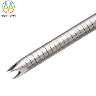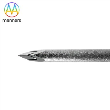


Cobalt Chromium Franseen Needles
Product Specification Product Show Patients who go to the hospital for abdominal ultrasound, especially for pancreatic ultrasound, often have to be prepared in advance, that is, take laxatives to expel the gas in the intestine, which can improve the quality of examination. Still, sometimes the...
Description
Product Specification
Product name | Cobalt Chromium Franseen needles |
material | Stainless steel, etc. |
Needle Size | 12G, 14G, 16G, 18G, 19G, 20G, 21G, 22G, 23G, 25G, 26G, 30G, 31G, 32G |
Properties | Injection & Puncture Instrument |
Custom feature | According to your 2D/3D Drawing or sample provided |
Package | Standard carton or according to customer's requirement |
Certification | ISO9001:2015, SGS |
Product Show
Patients who go to the hospital for abdominal ultrasound, especially for pancreatic ultrasound, often have to be prepared in advance, that is, take laxatives to expel the gas in the intestine, which can improve the quality of examination. Still, sometimes the results are not good enough. Endoscopic ultrasound was originally developed to overcome the shortcomings of in vitro ultrasound (B ultrasound) in diagnosing pancreatic lesions. Endoscopic ultrasound is a device equipped with a high-frequency ultrasound probe at the front of the endoscope. As the endoscope enters the lumen of the esophagus, stomach and intestine, the gastrointestinal wall and its adjacent structures and organs, such as the pancreas and bile duct, can be examined by ultrasound at the same time of endoscopy, and high-resolution images can be obtained. At present, there are two types of endoscopic ultrasonography for the examination of pancreatic diseases. One is that the front end of the endoscope is directly equipped with a high-frequency ultrasound probe, which can explore the entire structure of the pancreas in the duodenum and stomach. The other is through duodenoscope, the ultrasound probe is inserted through the endoscopic biopsy hole, extending from the front end of the scope, and inserted into the pancreatic duct from the opening of the duodenal papilla. The lesions in the pancreas can be intuitively detected, which is called "ultrasound pathological image". Endoscopic ultrasound can provide images of local lesions of the pancreas, especially for small tumors (such as pancreatic exocrine and endocrine tumors), which is superior to CT, ERCP and MRI to some extent.


Our Factory


Our Certification


Cobalt Chromium Franseen needles
Hot Tags: cobalt chromium franseen needles, China, suppliers, manufacturers, factory, customized, custom, cheap, low price, made in China