The use process of arthroscopy for arthroscopy
Jan 05, 2022
The miniaturization of arthroscopy has made it possible to simplify the operation to be performed in outpatient and local anesthesia, but we still use ordinary knee arthroscopy in the operating room. The steps are briefly described as follows.
Under continuous epidural anesthesia, place the affected limb on the operating table, use an epidural needle to perform joint puncture on the suprapatellar capsule, extract the exudate, and inject saline into the joint to expand the joint cavity (saline bottle hanging height Generally, it is about 1m higher than the knee joint). The puncture point is selected on the center of the triangle formed by the lateral edge of the patellar tendon, the anterior edge of the lateral malleolus of the femur, and the upper edge of the tibia. First make a small incision of about 0.5 cm in the skin, then puncture with a trocar matching the diameter of the arthroscope, remove the sharp obturator, replace it with a blunt obturator, and insert the arthroscope into the joint cavity. The observation sequence is as follows: Patella Upper synovial fold-patellofemoral joint-medial recess (internal inner wall, medial patellar synovial fold, recess surface of medial malleolus)-medial tibiofemoral joint (medial meniscus, front underside of femoral medial malleolus and opposite tibial articular surface )-And then to the suprapatellar capsule-lateral tibiofemoral joint (lateral meniscus, femoral lateral malleolus anterior and lower and opposite cavity bone seal writing)-lateral recess (lateral inner wall, recess surface of the lateral femoral condyle, muscle health). What you see above can be photographed. Finally, it can be used for biopsy. After washing, drain the full liquid, pull out the trocar, and suture the skin incision. Regarding different opinions on whether to use hemostasis during arthroscopy, the author believes that it is better not to use tourniquet at the beginning of the microscopic examination, so that the tissue structure in the joint can maintain a normal appearance, and it is easy to judge normal or abnormal tissue. For diagnosis, arthroscopic surgery (including membrane biopsy) is enough, about one hour of operation is enough; if you want to continue the therapeutic operation, you need to use a tourniquet.
Synovial pathological examination is an important step in diagnosing arthropathy. After the joint boundary examination is completed, synovial biopsy is usually performed at the same time. There are three methods for synovial biopsy: ①Blind examination: that is, when the arthroscopy is completed, the mirror is withdrawn from the sleeve, the biopsy forceps are inserted, the head of the biopsy forceps is felt through the skin with the other hand, and the biopsy is performed. ②If a special lesion area has been seen under the microscope, the original scope can be withdrawn from the casing, replaced with a small arthroscopy with biopsy forceps, and work under direct vision. ③If the surgeon wants to keep the observed lesions in the field of vision, he can insert a biopsy through the second puncture port for biopsy.
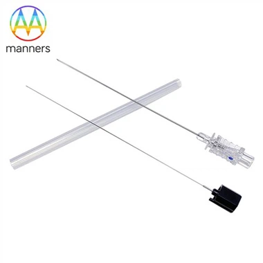
If you have any questions, please contact us. Our company can produce various customized needles, medical needles, puncture needles, hypodermic needles, biopsy needles, vaccine needles, injection needles, syringe needles, veterinary needles, pencil point needle, ovum pick up needles, spinal needles, etc. If you need customized needle products, please contact us. We look forward to your inquiry! The quality of the products manufactured in our factory will surely satisfy you!
Please contact us if you need: zhang@sz-manners.com