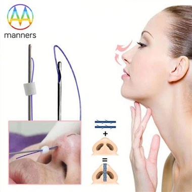
The route of the dural puncture needle
Dec 28, 2021
) Transsacral hiatus puncture method: The patient is placed in a prone position, and the skin is routinely disinfected. After local anesthesia, a 16-gauge epidural puncture needle is used to puncture the sacrococcygeal ligament at 45 degrees with the trunk, and then it is changed to 25 degrees to slowly penetrate into the sacral canal. Since the subarachnoid space ends at the second sacral plane, the puncture needle should not exceed this plane to ensure that it will not penetrate into the subarachnoid space, pull out the needle core, observe whether there is cerebrospinal fluid overflow, and insert the epidural anesthesia catheter Into the epidural space of the lumbosacral interstitial space, then the examination, the large site collection withdraws the puncture needle. Before injecting the contrast agent, you can inject 0.5% procaine 80-120ml first, and observe whether there is spinal anesthesia to rule out the dural penetrating injury. After confirming that there is no dural injury, the contrast agent can be injected. (2) Translumbar puncture and postdural radiography: the patient is placed on his side, the affected side is down, and his head is slightly raised. The method is the same as that of epidural anesthesia, and puncture is performed from the gap between waist 3 and waist 4. After confirming that the needle tip is located in the epidural space, the contrast agent is slowly injected, and then the puncture needle is removed and X-rays are taken. (3) Translumbar puncture and predural angiography: the patient takes the lateral position, and uses a 22-gauge puncture needle to penetrate between the lumbar 4 and lumbar 5 spines. When the needle tip breaks through the posterior arachnoid wall and enters the subarachnoid space, there is an outflow of cerebrospinal fluid, and the puncture continues forward. When the needle tip penetrates into the predural space, there is no more cerebrospinal fluid outflow. Take X-rays to confirm that the needle tip is at the posterior edge of the vertebral body. Inject 2ml of 0.5% procaine. If there is no resistance, inject 2ml of contrast agent. Then take X-rays to confirm that no contrast agent has spilled into the subarachnoid space. 4ml, pull out the puncture needle, it is complete. Epidural radiography can show the size of the spinal canal and the herniated disc outside the dural sac. It has a high accuracy rate for the diagnosis of lumbar disc herniation, but because the spinal epidural space has an unstable amount of fat, loose connective tissue and venous plexus, it affects the display of the spinal canal contour image. When reading the film, it is necessary to analyze the combination of positive and lateral positions.
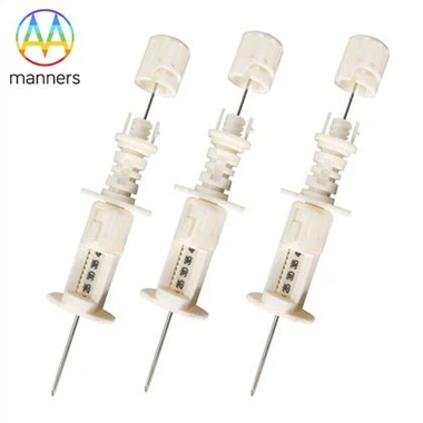
If you have any questions, please contact us. Our company can produce various customized needles, medical needles, puncture needles, hypodermic needles, biopsy needles, vaccine needles, injection needles, syringe needles, veterinary needles, pencil point needle, ovum pick up needles, spinal needles, etc. If you need customized needle products, please contact us. We look forward to your inquiry! The quality of the products manufactured in our factory will surely satisfy you!
Please contact us if you need: zhang@sz-manners.com