The level of forehead top occipital area and characteristic?
Oct 21, 2022
From the top of the cranium comes the superior orbital margin, and from the top comes the superior nial line and the external occipital carina. On both sides, the superior temporal line and the temporal region are demarcated.
1. Level: Soft tissue covering this area can be divided from shallow to deep
There are 5 layers of skin, superficial fascia, cap aponeurosis, fronto-occipital muscle, subaponeurotic tissue and skull outer membrane. The three layers of the superficial part are closely connected and not easy to separate, so it is always called scalp.
(1) Skin: The skin in this area is thick and dense, containing a large number of hair follicles, sweat glands and sebaceous glands, which are prone to furuncle or sebaceous cyst. Blood vessels and lymphatic vessels are also extremely rich, bleeding in trauma, but wound healing faster.
(2) Superficial fascia: composed of dense connective tissue and adipose tissue, the connective tissue forms many vertical trabeculae and divides the adipose tissue into small compartments, which contain blood vessels and nerves. Therefore, when there is infection in the superficial fascia, the exudate is mostly limited to the local area, not easy to spread, and the nerve endings are easy to compress and produce severe pain. In trauma, the broken end of the blood vessel is not easy to contract, so it is necessary to pressure hemostasis or suture hemostasis.
(3) Cap apONeurosis and fronto-occipital muscle: The aponeurosis is tough and dense, and it extends the fronto-abdomen before and the occipital abdomen after. Both sides gradually became thinner and continued in the superficial layer of temporal fascia. When the scalp was transverse lacerated and the aponeurosis was injured, the wound was dehiscence due to the retraction of the fronto-abdomen and occipital abdomen. When stitching the scalp, the aponeurosis should be sewn to reduce the tension of the skin and facilitate wound healing.
(4) Subaponeurotic loose tissue: It is the loose tissue layer between the cap aponeurotic membrane and the skull membrane, also known as the subaponeurotic space. This gap is extensive at the top of the skull, reaching anteriorly to the orbitals and posteriorly to the upper parietal line. There are several catheters communicating with the intracranial venous sinus in the space, so when infection occurs, it can spread to the intracranial through the guide vessels. When bleeding in this gap, large hematoma is often formed, and ecchymosis may appear subcutaneously in the upper eyelid.
(5) Skull outer membrane: thin and dense, connected with each bone by connective tissue, so it is easy to peel off during the operation. However, in the bone seam, it is closely healed with the intersuture ligament, so the subperiosteal hematoma is limited to the scope of one skull, and it is easy to distinguish from cap-like subaponeurotic hematoma.
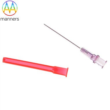
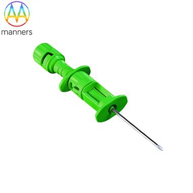
If you have any questions, please contact us. Our company can produce various customized needles, medical needles, puncture needles, hypodermic needles, biopsy needles, vaccine needles, injection needles, syringe needles, veterinary needles, pencil point needle, ovum pick up needles, spinal needles, etc. If you need customized needle products, please contact us. We look forward to your inquiry! The quality of the products manufactured in our factory will surely satisfy you!
Please contact us if you need: zhang@sz-manners.com