Examination of osteosarcoma
Sep 24, 2022
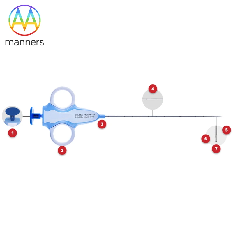
The EXAMINATION OF OSTEosARCOMA SHOULD FOLLOW 3 PRINCIPLES COMMONLY NAMELY OSTEosARCOMA EXAMINATION, THE FIRST step IS to WANT TO COLLECT CLINICAL DATA. First of all, the patient's age, the location of the disease, the location of the tumor, the symptoms, the mass examined, including the size and detection of whether there is muscular atrophy, whether there is compression sign, etc. After the completion of the clinical examination, imaging studies should be improved and the imaging examination including X-ray, is ordinary X-ray, shooting is a side of the lesion site, and even some special position, observe the presence of bone destruction, is there a periosteal reaction, namely common Codman triangle, these are some manifestation of osteogenesis sarcoma, and some specific signs of malignant tumor, Specific imaging findings. We do a CT scan. This CT scan can even be reconstructed in three dimensions under different conditions, such as cartilage tissue layer and bone bed, to observe the size and location of the tumor, the degree of bone destruction, whether it has broken through the bone cortex, and whether it has a soft tissue mass. If necessary, imaging enhancement can be done to see the blood supply of the tumor. The information provided by enhanced CT is very rich. Nuclear magnetic examination can see whether the infiltration into the soft tissues, infiltration into the surrounding vascular nerves, and even there is no edema around. Because MRI is an aqueous image and is very sensitive, it is sometimes used as a comparison of efficacy before and after chemotherapy. Other examination PET-CT, bone scan, according to the situation to choose. Will TELL TO osteosarCOMA, MUST HAVE THE EXAMINATION OF LUNG, INCLUDE CHEST PIECE AND ALVEOLAR LAYER CT, CHEST PIECE AND alveOLAR LAYER CT IS THE METASTASIS THAT DOES NOT HAVE LUNG EXCEPT, THE LESION THAT DOES NOT HAVE LUNG. When imaging is complete, laboratory tests, or laboratory tests, are performed. The patient of osteosarcoma often has alkaline phosphatase heighten, have the circumstance of calcium phosphorus metabolism even. At the same time, some differential diagnosis, such as myeloma may appear in the increase of M protein, urine may also have Ben's protein, serum immunoglobulin in the blood may appear abnormal M protein, this is some differential diagnosis. Increased alkaline phosphatase and calcium and phosphorus metabolism can also be used to make another differential diagnosis. For example, hyperparathyroidism, there will be an abnormal increase in alkaline phosphatase and high calcium, low phosphorus, and even bone destruction. This test also includes other tests, such as blood test, erythrocyte sedimentation rate, and C-reactive protein. After the imaging and laboratory tests are completed, the third step is to take a tissue biopsy. In general, needle biopsy, which is used to obtain the diseased tissue, is used for pathological biopsy. When the pathological examination is complete, it indicates that the diagnostic step of the patient has passed, and the next step should be to the treatment stage. It should be pointed out that the puncture site should be selected at the incision of the next operation, so that the puncture path can be removed at the same time. So the choice of puncture site should be made by the doctor.
If you have any questions, please contact us. Our company can produce various customized needles, medical needles, puncture needles, hypodermic needles, biopsy needles, vaccine needles, injection needles, syringe needles, veterinary needles, pencil point needle, ovum pick up needles, spinal needles, etc. If you need customized needle products, please contact us. We look forward to your inquiry! The quality of the products manufactured in our factory will surely satisfy you!
Please contact us if you need: zhang@sz-manners.com