Why Echo Tip Matters In OPU
Oct 07, 2026
The Hidden Risk of Losing the Needle Tip
Transvaginal oocyte pick-up (TV-OPU) is, by its very nature, an indirect procedure. The operator's hands hold a probe; the needle travels inside a fixed guide channel; and the only window into the pelvic cavity is the grayscale ultrasound fan. Unlike laparoscopic surgery, there is no direct line of sight, no tactile confirmation of tissue layers, and no ability to "see" the tip once it has crossed the vaginal mucosa. In routine anatomy this is manageable. But in the real world of reproductive medicine, routine is the exception. Patients arrive with retroverted uteri, previous cesarean scar niches, mild endometriosis creating peri-ovarian adhesions, body-mass indices above 30, or ovaries that have been pulled posteriorly by prior pelvic inflammatory changes. In these cases, a conventional polished stainless-steel needle behaves almost like a mirror: it reflects the ultrasound beam away from the transducer, producing either a faint thin line or, worse, a clean acoustic shadow that gives no positional information. The consequence is a dangerous cognitive loop: the tip is invisible, the operator assumes it is "just a little deeper," and applies more axial force. This is precisely how asymptomatic hematomas, incidental vessel nicks, and rarer bowel-wall contacts occur. A 2021 multicenter survey of European IVF units noted that in cases flagged as "difficult OPU," loss of tip visibility was cited as the single most common intraoperative stressor, ahead of even ovarian mobility. For the patient, this translates into longer procedure time, higher analgesic requirement, and a longer recovery window. For the embryology team, it means unpredictable follicular collapse and occasionally fragmented cumulus retrieval.
How Echo Tip Marking Works
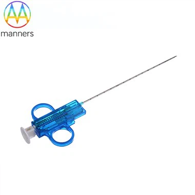
The physics of ultrasound contrast is the foundation here. An ultrasound image is built from returning echoes; smooth, specular surfaces reflect sound away, while micro-irregular surfaces scatter echoes back toward the probe. Echo tip marking deliberately engineers that scatter. The most common method is laser micro-etching: a ring or series of micro-grooves is ablated around the distal 3–5 mm of the needle, creating controlled roughness that returns a bright, persistent hyperechoic dot. Alternative approaches include sandblasted bands, titanium-nitride micro-coatings with differing acoustic impedance, and embedded polymer marker rings. The key design constraint is that the marker must not compromise lumen patency or outer diameter - the inner wall must remain polished to protect the cumulus–oocyte complex, while only the outer surface is textured at defined bands. On a 5–7 MHz transvaginal probe, a well-designed echo tip appears as a crisp point with minimal comet-tail artifact, allowing the operator to judge not just depth but also the angle of entry relative to the follicle wall. Some advanced designs add a second marker 5 mm proximal to the tip, so that the inter-marker distance on screen becomes a depth ruler: if the two dots appear 5 mm apart on the frozen image, the operator knows the insertion depth with sub-millimeter confidence.
Needle Variants in the Market
The current market offers a surprising variety. Laser-etched single-ring tips are the workhorse: durable through sterilization, visible on most mid-range probes, and cost-effective. Dual-echo band needles add a proximal marker for depth estimation and are favored in teaching hospitals. Coated echo tips using roughened titanium or ceramic micro-layers produce a brighter signal on low-frequency imaging, useful in obese patients, though coating wear over multiple passes is a consideration. Full-shaft graduated echo needles print faint echo bands every 5 mm along the shaft - popular for resident training but slightly noisier on screen. Bevel-orientation markers etch a small notch on one side of the bevel so the surgeon can rotate the cutting edge away from suspected vessels. Finally, curved-tip echo needles for high-fixed ovaries combine a pre-angled distal segment with an echo band at the apex, reducing the need for extreme probe tilting.
Operating Rules That Reduce Complications
The first rule taught in good OPU rooms: never advance a needle you cannot see. If the echo dot fades, freeze the image, rotate the probe 10–15°, or switch to power Doppler to re-localize. Second, respect the guide channel angle - do not "freehand bend" the needle out of plane; this is where lateral tissue tearing begins. Third, use short, deliberate thrusts of 2–3 mm rather than a single long push; this lets the capsule yield cleanly. Fourth, when a follicle fails to yield fluid, withdraw 1–2 mm inside the ovary and re-angle slightly before considering a second vaginal puncture. Fifth, always scan the pouch of Douglas at the end for free fluid or blood before removing the probe. These rules sound simple, but they are the difference between a 6-minute uneventful OPU and a 20-minute struggle.
What Clinics Learn Over Time
Senior embryologists and physicians develop an almost intuitive relationship with the echo dot. One veteran reproductive surgeon describes it as "watching a comet cross the screen." In internal audits shared at the 2023 ESHRE workshop, two centers that switched from plain to echo-tip needles reported a 30–40% reduction in junior-operator time-to-first-follicle and a measurable drop in post-OPU hematoma incidence, albeit from a low baseline. Nurses report fewer requests for rescue analgesia. Perhaps the most telling feedback: trainees say they "stop guessing" - and guessing is the enemy of safe OPU.
Summary
Echo marking is not a cosmetic feature stamped on a needle. It is a translation layer between steel and sound, converting a semi-blind pelvic puncture into a visualized, measurable trajectory. In an era where patients demand gentler, faster, more transparent procedures, that visibility is clinical value, not marketing.
Outlook
The next five years will likely fuse echo marking with digital guidance. Prototype systems already overlay a virtual needle path on the ultrasound screen by tracking the echo signature in real time. Coupled with AI vessel-segmentation on Doppler, the system can flash a warning when the projected path approaches an iliac vessel. The echo tip, once a simple etched ring, becomes the anchor point for augmented-reality OPU.