The Insight Into Structure: How Bone Marrow Biopsy Unravels The Spatial Code Of Hematological Diseases
Apr 14, 2026
The Insight into Structure: How Bone Marrow Biopsy Unravels the Spatial Code of Hematological Diseases
Q&A Approach
When a bone marrow aspirate smear shows a blurry cellular background, how can we determine if this reflects a universal state of the entire marrow or an accidental sampling of a focal lesion? Why do some blood diseases appear "calm" on smears yet reveal just "the tip of the iceberg" in biopsy sections? The significance of the bone marrow biopsy lies in its provision of the physician's first-ever ability to observe the "three-dimensional structure" of the marrow.
Historical Evolution
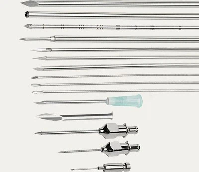
The cognition regarding bone marrow structure has undergone a paradigm shift from "cell suspension" to "tissue section." Before the 1950s, marrow diagnosis relied entirely on smears, often failing due to "dry taps" or dilution. In 1962, Burkhardt invented the first biopsy needle capable of retrieving intact marrow tissue, albeit samples were small and fragile. The 1971 Jamshidi needle, with its bevel design and inner cannula technique, made acquiring 1–2 cm intact cores feasible. The popularization of plastic embedding in the 1980s made thin sections (2–3 μm) and high-quality staining a reality. In the 21st century, the integration of digital pathology and artificial intelligence is bringing the interpretation of bone marrow structure into the era of quantification and intelligence.
Technical Standard Definitions
A qualified bone marrow biopsy core is a "biochip" carrying structural information:
|
Core Parameter |
Gold Standard Definition |
Clinical Value |
|---|---|---|
|
Length |
≥1.5 cm |
Ensures inclusion of at least 3–5 complete bone marrow units (BMUs). |
|
Diameter |
1.5–2.0 mm |
Provides sufficient field of view while minimizing trauma. |
|
Integrity |
No crushing, fracture; trabecular structure continuous |
Avoids artificial artifacts, reflecting true spatial conformation. |
|
Staining Req. |
H&E + Reticulin (Iron stain if needed) |
H&E assesses cell morphology/distribution; Reticulin shows fibrosis grade. |
The Four Dimensions of Structural Assessment
How a biopsy section is "read":
Cellularity Assessment
Method:At 100x magnification, assess the relative areas of hematopoietic tissue, adipose tissue, and bone trabeculae.
Quantitative Standard:In normal adult iliac bone, hematopoietic tissue accounts for ~30–70% (decreases with age).
Clinical Significance:Distinguishes Aplastic Anemia (hematopoietic tissue <20%) from Myeloproliferative Neoplasms (often >80%).
Cellular Spatial Localization
Normal Pattern:Granulocytic series near trabeculae; erythroid and megakaryocytes in the central marrow.
Abnormal Pattern:Clusters of blasts "back-to-back" against trabeculae (ALIP phenomenon)-a key diagnostic clue for Myelodysplastic Syndromes (MDS).
Stroma & Fibrosis Assessment
Reticulin Grading:Uses the European Consensus from MF-0 to MF-3 to quantify fibrosis.
Clinical Correlation:MF-2/3 grades correlate with Primary Myelofibrosis or fibrosis secondary to other marrow neoplasms, indicating poorer prognosis.
Vascular Structure & Bone Turnover
Observation Points:Increased vessel count or sinusoidal dilatation suggests tumor infiltration; abnormal resorption or thickening of trabeculae hints at metabolic bone disease or metastatic carcinoma.
Clinical Application: Filling Six Blind Spots of Smears
Biopsy holds irreplaceable value in these scenarios:
Investigating "Dry Tap": Differentiates between "packing" due to extreme hypercellularity versus fibrosis or hypocellularity.
Capturing Focal Lesions: Lymphoma, metastatic cancer, and granulomas (e.g., TB) are often focal; blind aspiration misses them, while biopsy dramatically increases detection rates.
Diagnosis & Grading of Fibrosis: An impossible task for smears; reticulin-stained biopsy is the gold standard.
Identifying Bone Marrow Necrosis: Biopsy reveals dissolved cell structures with an eosinophilic background, a sign of aggressive tumors or severe infection.
Assessing Residual Disease Post-Therapy: Post-chemotherapy marrow may show a "patchy" recovery; biopsy evaluates if lesions are replaced by normal tissue.
Accurate Iron Stores Assessment: Prussian Blue staining on biopsy visualizes storage iron within macrophages, diagnosing iron deficiency or overload more accurately than smears.
Future: From Morphology to Digital
Bone marrow structural analysis is undergoing digital transformation:
Whole Slide Scanning & AI Quantification: AI algorithms automatically calculate hematopoietic area, cellular density, and fibrosis ratio for objective, repeatable assessment.
Spatial Transcriptomics: Analyzing gene expression while retaining tissue location information, linking structure to function (e.g., profiling ALIP foci).
3D Marrow Reconstruction: Based on serial sections, reconstructing the 3D structure of micro-marrow cavities for a realistic simulation of the hematopoietic niche.
Conclusion
"Bone marrow biopsy allows us to see the 'forest' of the marrow for the first time, not just the individual 'trees.'" It is precisely this insight into spatial structure that makes it an indispensable tool for the precise diagnosis of hematological diseases, especially those adept at "hiding" or presenting as "focal" lesions.