The Engineering Marvel Of Thin-Wall-Large-Core Technology
Jul 24, 2026
Maximizing Breast Biopsy Yield Without Increasing Trauma
https://www.mayoclinic.org/tests-procedures/breast-biopsy/about/pac-20384812
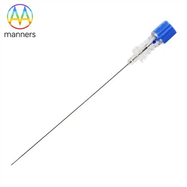
In the realm of minimally invasive breast excision and Core Needle Biopsy (CNB), surgeons constantly face a dilemma: they must obtain sufficiently large tissue samples to satisfy the demands of pathological typing and molecular testing, yet they must minimize puncture diameter to reduce risks of hemorrhage, hematoma, and breast disfigurement. The Thin wall for larger core technology employed in our Breast Biopsy Needle is an engineering masterpiece designed to solve this puzzle. By pushing the limits of needle tube geometry, this technology maximizes the effective area of the internal sampling channel without altering the external trauma footprint.
Traditional biopsy needle walls are thick, occupying valuable cross-sectional area. For instance, a standard 14G biopsy needle has an outer diameter of approximately 2.1mm. Under conventional manufacturing processes, maintaining the rigidity and kink resistance of the needle tube necessitates a greater wall thickness, severely limiting the actual available inner lumen diameter (i.e., the sampling notch width). Thin-wall technology revolutionizes this through improved seamless steel tube drawing processes. We utilize a specialized cold-drawing die, coupled with inline annealing treatments, allowing the needle wall to be thinned by 15-20% while retaining-and often exceeding-original standards for bending strength. This translates to nearly a 30% increase in sampling notch volume within the same 14G outer diameter.
This volumetric increase holds revolutionary significance for breast pathological diagnosis. Confirming a breast cancer diagnosis often relies on observing tissue architecture, particularly in invasive ductal carcinoma, where evidence of tumor cells breaching the basement membrane into the stroma is required. Insufficient sample size may prevent pathologists from capturing critical infiltration evidence, leading to underestimated tumor staging. Moreover, with the advent of precision medicine, genetic tests (such as Oncotype DX) require sufficient DNA/RNA extraction from paraffin-embedded tissues. The ample samples yielded by thin-wall-large-core technology enable a single needle pass to satisfy多重 requirements for H&E staining, immunohistochemistry (ER/PR/HER2), and FISH testing, eliminating the need for repeat punctures due to sample insufficiency and significantly alleviating patient physical and psychological burdens.
Achieving this technology hinges on precision Machining Technology. The wall-thinning process is not mere physical cutting but a complex material rheology process. We employ a multi-pass drawing process, with each draw followed by a bright annealing treatment. This eliminates internal stresses induced by cold working, restores material plasticity, and prevents cracking during subsequent processing. Simultaneously, to ensure absolute smoothness of the inner lumen, we use specialized oil-based lubricants during drawing, followed by ultra-precision cleaning to remove any residue. The smoothness of the inner wall directly impacts the ease of tissue core ejection; any microscopic burrs could cause tissue adhesion, resulting in sample fracture or "dry taps."
In needle tip geometry, thin-wall technology also presents new challenges. A thinner-walled tip is more susceptible to rolling or collapsing under high-speed impact. To this end, our Breast Biopsy Needle features a patented Precision grinding process. Using 5-axis CNC grinders, we form a unique double-bevel or triple-bevel structure at the needle tip. This design disperses insertion force across multiple vectors, reducing unit-area pressure and allowing the thin-walled tip to penetrate dense Cooper's ligaments smoothly. Concurrently, the ground cutting edge undergoes microscopic inspection to ensure edge radius is controlled at the micron level, achieving razor-sharpness. This sharpness not only reduces insertion pain but, crucially, facilitates "cutting" rather than "crushing," maximally preserving the original morphology of tissue cells.
Additionally, to complement thin-wall-large-core technology, we offer expanded Custom feature options. Based on customer-provided 2D/3D drawings, we can adjust the length and depth of the sampling notch to suit various biopsy gun (Biopsy Gun) models. For instance, Vacuum-Assisted Biopsy (VAB) systems require longer notches to ensure effective negative pressure suction, whereas manual biopsy needles benefit from deeper grooves to accommodate tissue. We can also apply hydrophilic coatings to the needle surface to further reduce friction coefficients, aiding the thin-walled needle in sliding more smoothly to the target location.
In conclusion, Thin wall for larger core technology is a crystallization of modern material science and precision manufacturing. It not only enhances the sampling efficiency of the Breast Biopsy Needle but also embodies the core tenet of minimally invasive medicine: achieving maximum benefit with minimum trauma. As 3D printing technology finds broader application in mold making, we anticipate topological optimization of the internal needle lumen, potentially breaking the wall-thickness barrier further while maintaining strength, thus providing more powerful tools for the early and precise diagnosis of breast diseases.