Technological Evolution And Future Trends—Tuohy Needles In The Era Of Smart Sensing And Visualization
Jul 22, 2026
Medical innovation remains relentless. The venerable Tuohy needle, while conceptually stable for decades, now stands at a transformative juncture. Converging advancements in materials science, microelectronics, and image guidance are catalyzing an evolution from purely mechanical puncturing tools toward intelligent, visualized interventional platforms. As industry stakeholders, prognosticating these trajectories is vital for steering future R&D and manufacturing strategies.
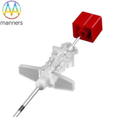
The most conspicuous shift involves visualization technologies. Traditional epidural access ("blind technique") hinges heavily on tactile feedback and experience. However, anatomical variations (obesity, scoliosis, prior surgery) challenge even veteran practitioners. Ultrasound-guided regional anesthesia (UGRA) alters this paradigm. Modern Tuohy needles now incorporate echogenic enhancements. Laser-etched micro-grooves, helical patterns, or sandblasted finishes modify acoustic interfaces. Instead of appearing as fleeting hypoechoic lines, needles manifest as vivid "comet-tail artifacts" or continuous hyperechoic dots under ultrasound. This physical modification-requiring no structural alteration-dramatically elevates needle tip visibility and placement accuracy, proving invaluable in pediatric or bariatric populations.
Deeper innovation resides in smart sensing integration. Research frontiers explore embedding micro-sensors within needle tips or walls. Optical Coherence Tomography (OCT) micro-probes could furnish real-time, micron-resolution cross-sectional tissue imaging during advancement, differentiating ligamentum flavum, epidural fat, and dura mater-surpassing subjective "loss of resistance" interpretation. Miniature pressure transducers represent another avenue. Monitoring real-time interstitial fluid pressure offers objective data: a characteristic pressure drop upon transitioning from high-resistance ligamentum flavum into the low-pressure epidural space. Fiber Bragg Grating (FBG) sensors multiplex strain, temperature, and pressure sensing, potentially detecting contact with neural structures and triggering millisecond alerts to preempt nerve injury.
Materials science crossovers portend further possibilities. While 304/316 stainless steel excels mechanically, its rigidity can prove disadvantageous in tortuous anatomy. Nickel-Titanium (Nitinol) alloys, possessing shape-memory and superelastic traits, are emerging for specialized applications. Nitinol needles might remain straight externally but assume pre-programmed curvatures upon reaching body temperature, navigating around bony spurs or vessels to reach targets. Biodegradable polymers, though currently unsuitable for rigid Tuohy needles, warrant exploration for short-dwell guidewires or specialized scaffolds, potentially eliminating removal procedures.
Human-Computer Interaction and Virtual Reality (VR) training constitute another domain. High-fidelity simulators, incorporating haptic feedback devices replicating ligamentum flavum resistance, immerse trainees within virtual anatomy. VR headsets overlay realistic visuals. Trainees practice Tuohy needle insertions repeatedly, receiving instant scoring on trajectory, angulation, and force dynamics. This curtails clinical learning curves and mitigates patient risk during skill acquisition. Manufacturers contribute by supplying authentic needle biomechanical data to refine simulator physics engines.
Looking ahead, Tuohy needle evolution will likely transcend the "needle-as-tool" concept, embracing "Needle + Imaging + Data" ecosystems. Future iterations might house miniature CMOS cameras transmitting live video feeds during insertion. Artificial intelligence algorithms could analyze impedance or imaging data, autonomously identifying tissue planes and recommending optimal trajectories. Targeted drug-eluting capabilities might activate immediately upon confirmed positioning. While bordering on science fiction today, such integrations progress steadily via micro-electromechanical systems (MEMS) and nanotechnology. We remain committed to R&D investment, tracking these frontiers to translate breakthroughs into safe, effective clinical solutions-ensuring this classic invention retains vitality in the modern era.