Technical Integration And Optimization Of Stereotactic Systems And Biopsy Needles
Jun 14, 2026
https://my.clevelandclinic.org/health/diagnostics/24204-breast-biopsy-overview
Core Perspective: Analyzing how stereotactic systems (such as X-ray, MRI, and Ultrasound) synergize with different types of biopsy needles (such as spring-loaded cutting needles and vacuum-assisted biopsy probes) from an engineering and device integration standpoint to achieve optimal performance.
Breast stereotactic core needle biopsy is not a solo performance by a single device, but a concerto played by a sophisticated system. This system comprises imaging equipment, positioning devices, biopsy guns, and biopsy needles. The seamless integration between these components is the key to ensuring operational safety, efficiency, and accuracy.
First, the imaging modality determines "what to see." X-ray stereotactic systems excel at visualizing microcalcifications and are the preferred choice for biopsies following mammographic screening. Their principle involves calculating the 3D coordinates of a lesion via dual exposures at ±15° angles. In contrast, MRI stereotactic systems offer exceptionally high soft tissue resolution, clearly delineating occult lesions, making them particularly suitable for high-risk populations. However, MRI systems impose strict compatibility requirements on biopsy needles-only non-ferromagnetic titanium alloy or specialized polymer needles may be used, as ferrous metals would cause severe safety hazards and image artifacts.
Second, the positioning device dictates "how to get there." Whether utilizing a horizontal platform or a vertical biopsy chair, the core function is to use high-precision robotic arms or electric motors to deliver the biopsy needle tip precisely to the calculated coordinates. Modern systems are universally equipped with "coaxial sheath" technology: a larger bore introducer sheath is placed first to establish a channel, through which the biopsy needle can be passed repeatedly for multi-site sampling. This approach minimizes tissue trauma and reduces compression of the lesion.
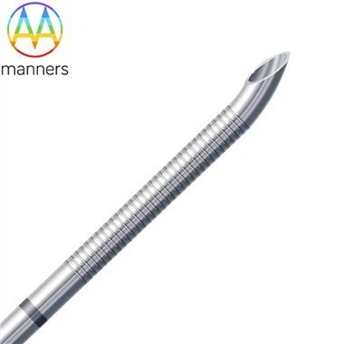
Finally, the type of biopsy needle determines "how much to take." Currently, two main types dominate the field:
- Spring-Loaded Cutting Needles: These utilize a spring mechanism to rapidly fire the inner stylet and outer cannula, instantly cutting and trapping tissue. The advantages are speed and simplicity, but they typically acquire only a single core per pass and may cause a pushing effect on hard lesions.
- Vacuum-Assisted Biopsy (VAB) Needles: These utilize negative pressure to draw tissue into the sampling notch, where a rotating blade then severs it. The advantages include continuous, multi-directional sampling, allowing multiple cores to be obtained via a single insertion. This results in a larger specimen volume and higher tissue integrity. These needles typically require connection to a dedicated console, which also manages irrigation and localization clip deployment.
Excellent system integration is manifested when the physician can view the real-time relative position of the needle tip to the lesion on the screen and control the vacuum pump and cutting action via a foot pedal. Some high-end systems even feature "smart sensing" capabilities, automatically adjusting the cutting force when the needle tip encounters dense fibrous tissue to prevent bending or breakage. In the future, integrating stereotactic systems with robotic arms promises to realize fully automated "one-click biopsies," further reducing human error.