Site Choice And Depth Control In Intraosseous Access
Nov 20, 2026
Why Site Selection Is a Physiological Decision
Site selection is usually taught as a matter of landmarks: find the tibial tuberosity, measure down, avoid the growth plate. Landmarks matter, but they are the surface expression of a deeper set of decisions. The site determines how much marrow is available, how stable the needle will be during transport, how much the device interferes with other resuscitation tasks, and how quickly a complication will be noticed.
Different sites behave differently under the same conditions. A site chosen for accessibility may be poorly perfused in shock. A site chosen for perfusion may be awkward to secure while a cervical collar is in place. A site that works well in a stationary patient may be the worst option during extrication. Recognizing these trade-offs is what separates a considered choice from a rote one.
Depth control is the second half of the same decision. The site defines the cortical thickness the needle must cross, and cortical thickness defines how far the tip can travel before it exits the far wall. Treating site and depth as a single combined problem is the most reliable way to avoid the complication that matters most: an apparently successful insertion that delivers fluid into soft tissue. It is also why the two are weighed together whenever a needle for intraosseous access is chosen for a service.
Comparing the Proximal Tibia, Humerus, and Distal Alternatives
The proximal tibia is the default for good reasons. The flat medial surface offers a broad target, the landmark is palpable even in patients with abundant soft tissue, the site is remote from the airway and thorax, and the needle is easy to secure without interfering with a collar or a pelvic binder. In adults the insertion point is roughly two centimeters below and slightly medial to the tibial tuberosity; in children the same relationship holds at a reduced distance.
The proximal humerus is the principal alternative. It is useful when the lower limbs are injured, trapped, or occupied by other equipment, and it offers a large medullary cavity in adults. Its disadvantages are practical: the site is closer to the thorax, less convenient during ongoing chest compressions, and harder to secure in a moving vehicle. It also requires a different landmark set, which means a team must train both sites if it intends to use both.
Distal options, including the distal tibia and, in young children, the distal femur, serve specific situations. They are useful when proximal sites are compromised by fracture, burns, or prior instrumentation. They carry a higher risk of needle dislodgement because the surrounding anatomy moves more, and they demand precise landmark identification, particularly in infants where the margin between the insertion point and the growth plate is small. Whatever site is chosen, the device's labeled indications and local protocol govern.
Depth Control: Cortex, Marrow, and the Far Wall
Depth errors occur in two directions. A needle that stops in the cortex produces no flow and a misleading resistance during flush. A needle that passes through the medullary cavity and out the far cortex produces free flow, a needle that may still stand, and extravasation that appears minutes later as swelling.
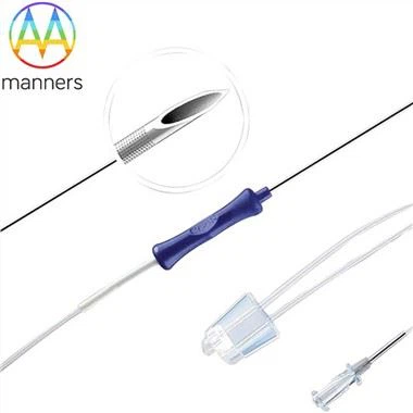
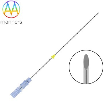
The end point that matters is the loss of resistance at the moment the tip enters the marrow. That signal is only useful if the operator is not applying so much force that the transition is masked. This is the structural argument for a cutting-tip intraosseous access needle driven by a low-power motor: because the tip machines a channel and the motor supplies the energy, the operator's hand is free to feel the change rather than to generate it. With minimal axial load, the passage from cortex to marrow is distinct, and the operator can stop within a millimeter rather than a centimeter.
Depth is also a function of needle length, which is why a single-length inventory is a depth-control hazard. A 15G stainless steel needle suits most adults, but the same length in a small child can reach the far wall with a single firm push. Systems that offer multiple lengths behind one driver allow length to be selected as a deliberate decision, and custom lengths produced to 2D or 3D drawings or to a physical sample allow a service to match unusual anatomy rather than compromise on it.
Preparing the Site and Seating the Needle
Preparation is short and consequential. Expose the site fully; a landmark palpated through clothing is a guess. Identify the tibial tuberosity by feel, then move distally along the flat medial surface. Cleanse with antiseptic and allow it to dry, since a wet site compromises both drape adhesion and dressing security.
Stabilize the limb against a firm surface rather than against another rescuer's hand. Position the needle perpendicular to the bone, without angling toward the joint or away from it. Activate the driver and apply light guidance only, letting the tip engage the cortex. When resistance falls away, release the trigger immediately and remove the driver while holding the hub steady.
Confirm before securing. Aspirate gently; marrow may or may not return, and its absence is not proof of failure. Flush a small volume against resistance and observe the site. Then confirm that the needle stands unsupported, which is the most practical evidence that it has passed the cortex and is seated in the cavity rather than resting in soft tissue. Apply the stabilization dressing, flush in pulses, and reassess after every patient movement.
Field Experience and Depth-Related Complications
The most instructive complication is extravasation. It presents as progressive firmness around the site, sometimes accompanied by a slow decline in flow rate. Because resuscitation produces edema independently, the finding is easy to attribute to the patient's condition rather than to the needle. Palpation at fixed intervals, rather than only when flow seems poor, is what catches it early.
The second complication is dislodgement, which is a depth problem in disguise: a needle seated shallowly has less shaft engaged in cortical bone and tolerates less torque. The third is failure to infuse, which is often attributed to a blocked needle and is more often a needle positioned against the far wall or in the cortex. Before replacing the device, reposition slightly and reassess; replacement costs time and adds a second puncture.
A recurrent field error is inserting at a site chosen for convenience during packaging or access rather than for perfusion and stability. Another is skipping the drying step for antiseptic, which produces a dressing that lifts during transport. Both are avoidable with a fixed sequence that is rehearsed rather than improvised.
What Correct Depth Buys
Correct depth produces more than flow. It produces stability, because a needle engaged through the full cortical thickness resists leverage. It produces predictable infusion, because the tip sits in the sinusoidal bed rather than against bone. It produces earlier recognition of complications, because a well-seated device with a secure dressing makes changes at the site visible rather than hidden.
Above all, correct depth produces confidence in the confirmation step. A clinician who knows what a properly seated needle feels like can distinguish a functioning line from a plausible one, and that distinction determines whether the next drug actually reaches the central circulation.
Emerging Practice and Specification Implications
Practice is converging on two principles: choose the site by physiology and access needs rather than by habit, and control depth with the device rather than with reaction time. Equipment is following, with shorter needle variants, clearer depth referencing, and stabilization systems that keep the hub from levering during transport.
For teams writing specifications, the questions are specific. Does the platform offer at least two lengths for the primary site, and can it accept custom lengths? Is the tip a cutting geometry paired with a low-power driver, so that the loss of resistance is felt rather than forced? Are the needles stainless steel with shafts that resist bending? Can the supplier produce to your 2D or 3D drawings or to a submitted sample, and does the standard carton packaging support site-specific kit configuration? A needle for intraosseous access is only as good as the depth at which it stops, and depth is decided by design as much as by hand.