Route Anatomy: Where The Needle Goes
Nov 05, 2026
The Pain Point
Vaccine failures are often blamed on the vaccine itself - and sometimes they are justified. But an equally common cause is invisible to the naked eye: the needle delivered the dose to the wrong tissue. The same vaccine, the same volume, and the same day can produce strong immunity in one flock and disappointing titers in another, simply because of where - and how deep - the injection landed. In poultry medicine, the route is a decision made in seconds but paid for over the entire grow-out cycle. Too shallow, and the dose leaks or sits in fat where immune cells are scarce; too deep, and the vaccine enters muscle or body cavities where absorption is erratic or tissue damage is severe. The anatomy of the injection site is the forgotten half of every vaccination protocol.
The Principle
Poultry skin and muscle are not uniform media. Subcutaneous space in the neck and thigh is a loose connective-tissue plane rich in lymphatic vessels and antigen-presenting cells - an ideal environment for slow-release vaccines, provided the dose is deposited in the plane itself and not in the dermis above it or the muscle below it. Intramuscular tissue offers a dense blood supply that absorbs vaccines quickly, but it also concentrates inflammatory responses; an oil-emulsion vaccine injected too shallowly into the muscle fascia creates granulomas and abscesses, while one injected too deeply may strike bone or major vessels. The wing-web route exploits a thin, well-vascularized membrane that responds rapidly to fowl pox and certain recombinant vaccines, but it demands precision that standard needles struggle to deliver. Each route has a preferred tissue plane, a safe depth window, and a tolerance for error - and the needle's length, bevel, and gauge are the instruments that determine whether the dose lands inside that window.
Equipment Classification
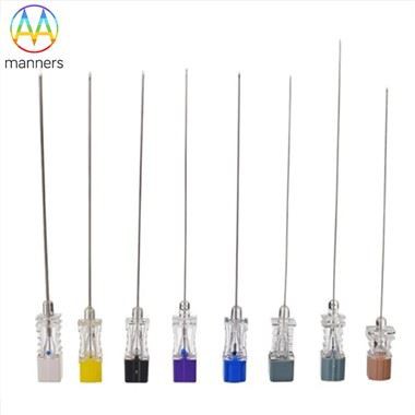
Route-specific needle design is a real category in poultry equipment. Subcutaneous needles are typically short - 12 to 18 millimeters - with a long bevel that creates a small entry wound and slides easily through the loose skin layer; gauge ranges from 20g to 25g depending on vaccine viscosity. Intramuscular needles are longer and sturdier, often 18 to 25 millimeters, with a short bevel and heavier wall to resist deflection when penetrating dense pectoral muscle; gauges of 16g to 20g are common for oil-emulsion products. Wing-web stab needles are thin and short - 22g to 25g - designed to pierce the membrane without transfixing the wing. In-ovo needles, used in hatchery automation, are ultra-precise, angled cannulas that deposit vaccine into the amniotic cavity of incubated eggs. Between these families, hybrid designs add depth-limiting collars or angle guides that convert guesswork into repeatable geometry.
Practical Guide
Map your protocol to anatomy before you map it to the calendar. For subcutaneous injection in the neck, pinch the skin to create a tent, insert the needle parallel to the body axis at a shallow angle, and confirm the tip moves freely under the skin before depressing the plunger - resistance means you are in the dermis. For intramuscular injection in the breast, insert perpendicular to the muscle surface, to a depth no greater than half the measured muscle thickness at that point; over-injection into the keel bone is the most common deep-error in broiler vaccination. For wing-web application, pierce the underside of the web and deposit the dose in the membrane itself, avoiding the large vessels visible through the skin. Calibrate needle length to bird age at every session - a 25 millimeter needle appropriate for a 35-day broiler is dangerous for a 10-day chick. And record the route, depth, and any injection-site reactions on the vaccination log, because the log is what turns experience into protocol.
Field Experience
A duck farm in Jiangsu consistently underperformed on Newcastle disease titers despite flawless vaccine handling and record-keeping. An independent audit found that operators were injecting the subcutaneous dose into the duck's neck muscle instead of the loose skin plane - the needle was long enough to pass through the tented skin and into the muscle beneath, delivering an intramuscular dose of a slow-release vaccine designed for subcutaneous uptake. The immune response was suppressed and local reactions were elevated. Retraining operators to shorten the needle exposure, tent the skin properly, and verify tip movement before injection restored expected titers within one cycle. The farm now includes a route-anatomy demonstration in every operator onboarding session.
Summary
The route is a decision with anatomical consequences. The same vaccine can immunize or irritate depending on the tissue plane it reaches, and the needle is the instrument that makes the difference. Farms that understand the anatomy of the injection site - not just the label on the vaccine bottle - convert vaccination from a routine task into a precision operation. Depth, angle, and tissue plane deserve the same documentation as dose and batch number.
Future Outlook
Depth-limiting needle technologies - collars, sheaths, and sensor-equipped cannulas that detect tissue resistance in real time - are moving from human medicine into veterinary equipment. Combined with ultrasound-validated anatomical charts for each breed and age, the next generation of poultry vaccination will replace "feel" with measurement. Route accuracy will become a logged parameter, auditable after every campaign, and the anatomy of the injection site will finally receive the engineering attention it deserves.