Real-Time Sonographic Visualization And The Art Of Needle Advancement in Ultrasound-Guided FNA
Jul 18, 2026
https://www.mayoclinic.org/tests-procedures/breast-biopsy/about/pac-20384812
With the rapid evolution of imaging technology, the traditional "blind" approach to FNA has transitioned toward precision procedures under real-time ultrasound guidance. This process tests not only the operator's hand-eye coordination but also represents a practical exercise in acoustic physics, where every angle and needle pass reflects profound technical expertise.
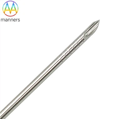
During ultrasound-guided FNA, the physician must meticulously calibrate probe frequency and applied pressure. Since 22G–25G needles are ultra-fine, their sonographic visibility pales in comparison to the bright, crisp delineation of a 14G core needle. The weak echo signals generated by fine needles are highly susceptible to deflection or bending artifacts induced by probe compression. Mastery of "needle tip visualization techniques" is therefore essential. By adjusting the incident angle-typically maintaining an optimal 30°–45°-the operator leverages the characteristic "comet-tail artifact" produced at the needle-tissue interface to track the tip precisely. Throughout the FNA process, minute hand tremors are amplified on the ultrasound screen. Thus, operators must establish a stable mechanical fulcrum, often anchoring the hypothenar eminence against the patient's chest wall to immobilize the wrist and ensure trajectory accuracy.
The artistry of needle advancement manifests profoundly in the real-time assessment of lesion consistency. During FNA, dynamic tactile feedback and sonographic changes allow differentiation between cystic and solid lesions. Upon entering a simple cyst, the operator perceives a distinct "pop" or loss of resistance, while sonography confirms immediate fluid aspiration and cavity collapse-often concluding within seconds. For solid nodules, the needle encounters subtle variations in traverse resistance. The operator performs rapid "piston-like" motions-quick in-and-out movements combined with intermittent suction. Given the fine caliber, negative pressure must be carefully titrated: excessive suction draws in diluting blood, while insufficient vacuum fails to retrieve adequate cellular material. This delicate calibration of force constitutes a core technical hurdle in FNA, typically requiring hundreds of supervised procedures to master.
Moreover, targeting microcalcifications presents significant technical challenges. The needle lumen is too narrow to reliably core out calcific particles as a coarse needle might. Instead, the operator often relies on a "vacuum aspiration effect," applying gentle, sustained suction to draw calcification-laden interstitial fluid into the hub. This demands exquisite sensitivity to subtle grayscale changes on ultrasound. Once confident in tip positioning within the target, the operator must swiftly aspirate and withdraw to minimize tissue trauma and bleeding risk. Although the entire FNA sequence is brief-typically 5–10 minutes-each step integrates the interventional radiologist's deep understanding of anatomy, physical principles, and refined motor skills.