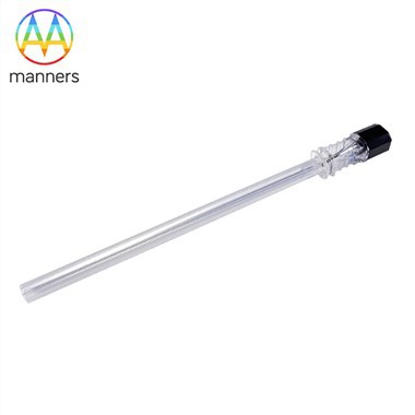
PTC Needle Guided Differential Diagnosis Between Intrahepatic Stone And Biliary Malignancy
Nov 11, 2026
1. Clinical Pain Points
Intrahepatic cholangiolithiasis and intrahepatic cholangiocarcinoma can both cause bile duct stricture and proximal duct dilation. In the stationary phase, stone patients have no obvious jaundice or fever, while early malignant lesions also show non-specific liver function abnormality. CT and B-ultrasound cannot reliably judge whether the stenosis edge is smooth or irregular. Misdiagnosis leads to delayed anti-tumor treatment or unnecessary liver resection for benign stone patients. ERCP often fails to reach high intrahepatic lesions. Without direct cholangiography roadmap, clinicians cannot tell the difference between benign annular stenosis caused by stone irritation and malignant infiltrative stenosis.
2. Working Principle
PTC needle delivers contrast medium into the upstream bile duct above stenosis. Under fluoroscopy, the contour of stenosis edge, the shape of duct dilation and filling defect are visualized. Benign stone-related stenosis usually shows smooth annular narrow segment, proximal spindle-shaped or dumbbell-shaped dilation, and clear stone shadow. Malignant stenosis shows irregular, long-segmented and serrated duct wall, and the dilation range is asymmetric. The stainless steel hollow needle provides stable injection channel, ensuring contrast fills the whole upstream duct without disturbance. The obtained X-ray features serve as the core basis for differential diagnosis, and can be combined with CT for comprehensive judgment of biliary cirrhosis and malignant transformation.
3. Classification of PTC Needles for Differential Diagnosis Scenarios
Fine diagnostic needles (0.25–1.2 mm): for patients with mild dilation and uncertain diagnosis, low trauma, suitable for initial differential screening. Medium dual-purpose needles (1.2–5 mm): for highly suspected malignant lesions, reserved for guidewire passage and subsequent biopsy or drainage. Custom special needles: customized tip geometry for hard-to-reach peripheral bile duct branches. Surface finish options: polishing, electroplating, plating and ultrasonic cleaning. ISO9001 certification is available. Diameter range 0.25–30 mm, customized by drawing or sample, packaging can be customized.
4. Operation Guide for Differential Diagnosis
Preoperative review: contrast-enhanced CT, liver function and tumor markers. Mark target dilated bile duct and suspicious lesion area under ultrasound. Select fine needle for first puncture. Confirm bile return before injecting contrast. Take multiple oblique fluoroscopy images to fully display the stenosis margin. Record whether the stenosis is short annular or long irregular, whether dilation is spindle-shaped or asymmetric, and whether filling defect is clear and mobile. After imaging, withdraw needle and compress the puncture site. If imaging features highly suggest malignancy, prepare subsequent interventional biopsy or drainage plan. If the imaging matches classic stationary intrahepatic stone signs, arrange conservative or surgical stone removal plan.
5. Practical Experience
Smooth annular stenosis with clear stone shadow is the most typical sign of benign intrahepatic cholangiolithiasis. When part of the bile duct fails to develop, operators need to distinguish it from malignant duct occlusion. Benign occlusion is usually caused by single large stone blocking the lumen; malignant lesions often show gradual tapering and irregular wall. For patients with biliary cirrhosis, the risk of concurrent cancer increases, so PTC and CT must be evaluated together. Operators should avoid over-injection pressure, which may cause duct wall tear and false irregular image interfering diagnosis. Many cases prove that PTC can correct the previous misdiagnosis of hepatitis or gastritis and identify early malignant lesions hidden behind stone lesions.
6. Summary
PTC needle enables direct visualization of biliary tree morphology, which solves the difficulty of differential diagnosis between benign stone stricture and malignant biliary obstruction. Different needle specifications match screening and high-suspicion scenarios. The X-ray signs captured by PTC form intuitive diagnostic evidence, which complements CT and laboratory indicators. Rational needle selection and multi-angle filming improve the accuracy of lesion nature judgment.
7. Prospect and Suggestion
Future PTC needle can integrate micro-biopsy channel to realize imaging and tissue sampling in one puncture. Manufacturers should maintain stable precision of fine needles for differential diagnosis. Radiology departments should build image libraries of benign and malignant biliary stenosis for physician training. For patients with unexplained intrahepatic duct dilation and negative tumor markers, PTC examination should be recommended to exclude early malignant lesions. Medical device suppliers need to provide fast customized service for special peripheral duct puncture needs.
< img alt="news-1-1" src="/uploads/24240/news/202604241539186d0ff.png " />