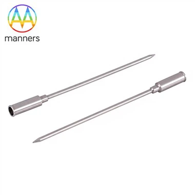
Nerve Block Puncture Needle Is Commonly Used in Clinical Anesthesia
Dec 01, 2022
It mainly includes nerve stem (ulnar nerve, femoral nerve, etc.), nerve plexus (brachial plexus, cervical plexus, etc.) and ganglion (stellate ganglion) block.
Common features are: injection of local anesthetics around the corresponding nerve (plexus), temporarily blocking the conduction of nerve impulses, temporary loss of sensation and/or motor function in the innervated area, analgesia and muscle relaxation for surgery, or pain treatment.
Due to the differences in anatomical location and distribution, the complications and adverse reactions caused by nerve (plexus) block in different parts are also different.
Because the location and course of nerves (plexus) are not easy to accurately locate through the body surface, puncture is mostly based on clinical experience, so the success rate of anesthesiologists with less experience is relatively low, and the incidence of complications is relatively high, especially in the case of repeated searching for different sensation and repeated puncture. It is more likely to cause damage to surrounding tissues, such as bleeding caused by damaged blood vessels, hematoma and compression of surrounding tissues and organs, or punctured pleura resulting in pneumothorax, lung damage, and local anesthetic poisoning and other serious complications.
In order to improve the success rate of nerve (plexus) block and reduce the occurrence of complications, many years of clinical exploration have been carried out, such as the selection of puncture points according to the surgical site during brachial plexus block, the high and low approach of the intermuscular sulcus, the subclavian approach, the subcoracoid process, the paracoracoid process, and the coracoid brachial space approach. Surface positioning of the first rib, multiple injection of one injection, and continuous or multiple block combination, etc. Cervical plexus block was performed by modified C4 transversal one-point method, high head puncture of intermuscular sulcus and so on. In recent years, the surface localization of nerve stem (plexus) with neurostimulator has been used in clinic and some experience has been gained. These methods are helpful to improve the success rate of puncture, but anesthesia complications still occur from time to time. The causes of complications include unskilled or wrong operation, improper choice of puncture approach and abnormal patient anatomy.
Anesthesia lessons of cervical plexus and brachial plexus blocks
The cervical plexus is composed of C1-4 spinal nerves, which congregate into the cervical plexus trunk under the prevertebral fascia after coming out of the foraminoforamen. At the midpoint of the posterior margin of the sternocleidomastoid muscle, a superficial plexus emerges, which innervates the skin sensation and muscle movement of the occipital and neck. Due to the very complex structure of the neck, some tissues of the larynx, trachea and floor of the mouth are not innervated by the cervical plexus. Cervical plexus block is most commonly used in thyroid surgery, and the nerve innervation of thyroid happens to involve sympathetic nerves and vagus nerves. Therefore, even though cervical plexus block is very perfect, it is difficult to meet the requirements of complete painless and comfortable thyroid surgery, nor can it completely inhibit the adverse reactions such as choking and heart rate change caused by surgical stimulation of the larynx and trachea.
The brachial plexus consists of the anterior branch of the C5~T1 spinal nerve, which mainly innervates the sensory and motor functions of the upper limbs and shoulders. Brachial plexus block is mainly used for upper extremity anesthesia and analgesia, but also for upper extremity Raynaud's disease or upper extremity vascular anastomosis vascular dilatation and other treatment. Brachial plexus block can be divided into single method or continuous method according to the administration method. According to different puncture approaches, it can be divided into three types: intermuscular sulcus, supraclavicular and axillary approach. Less commonly used methods are subcoracoid or subclavian triangle approach, etc.
Brachial plexus is adjacent to important structures such as internal jugular vein, common carotid artery, subclavian artery, vein and pleura, etc. Puncture injection into blood vessels can cause severe local anesthetic poisoning, puncture pleura and lead to pneumothorax or lung tissue injury.
The brachial plexus is located under the prevertebral fascia. There is normally a longitudinal connective tissue interval between the left and right prevertebral fascia, but in individuals this interval may not be complete and drugs injected from one side may enter the other side. The neck tissue is relatively shallow. When the puncture needle enters more than 2cm in brachial plexus block, it may penetrate into the spinal canal, leading to high epidural block or even spinal anesthesia and endangering life.