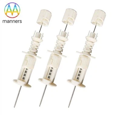
Laser Cutting And Marking On Marrow Biopsy Needles
Nov 19, 2026
Marks That Fade and Edges That Do Not Cut
Two complaints recur in needle quality review. The first concerns markings: depth graduations that become unreadable after the first wipe with an alcohol swab, or a lot code that cannot be resolved under the magnifier when a complaint is being investigated. The second concerns edges: a biopsy window whose lower lip tears the core instead of containing it, or a cut edge that carries a burr which sheds particulate into the marrow cavity.
Both are laser processes, and both are frequently specified in one line of a drawing with no process parameters attached. A supplier who receives "laser cut window, laser marked graduations" has been given an outcome, not a specification. The consequence is variability that only shows up in use, when an operator cannot read the depth at the moment it matters or a pathologist receives a core that has been shredded by an imperfect edge.
How the Beam Modifies Stainless
Laser cutting removes material by localised melting and vaporisation. In a thin-walled stainless cannula, the dominant concerns are heat input and assist gas. Excessive heat produces a recast layer and a heat-affected zone whose microstructure differs from the parent tube, and that zone is where corrosion and crack initiation begin. Insufficient assist gas pressure leaves dross on the inner wall, which narrows the lumen and can detach later as particulate.
Laser marking works by a different mechanism. Annealing marks produce a controlled oxide colour change with negligible material removal, and are the usual choice for depth graduations because they do not create a notch. Deep engraving produces a permanent, high-contrast mark but removes material and creates a site where crevice corrosion can start. The choice between them is a trade-off between readability and surface integrity, and it should be made explicitly for each mark on the device.
Classifying Laser Operations on a Marrow Needle
Laser operations on these instruments fall into four groups. Cutting of the biopsy window or side aperture determines core capture geometry and must be controlled for edge squareness and lip sharpness. Cutting and forming of the tip, where a laser is used rather than grinding, determines penetration behaviour and requires control of the recast layer at the bevel.
Marking of depth graduations is the third group, and it is the most clinically visible. Spacing, contrast and resistance to disinfection all matter, particularly where a single insertion depth has to be confirmed without a second look. The fourth group is identification marking: lot numbers, size codes and, increasingly, unit-level codes on the hub, which must survive sterilisation and remain legible for the life of the record.
For disposable marrow aspiration biopsy needles produced in a range of sizes, laser marking also carries a practical benefit. Marking the gauge and lot directly on the hub allows the device to be identified after the pouch has been opened, which is exactly when traceability is most often lost.
Setting and Verifying the Process
Specify parameters rather than intentions. For each cut, define the wavelength, pulse energy, repetition rate, focal position, assist gas and pressure, and the number of passes. For each mark, define the marking method, the target contrast value and the depth of any material removal. A drawing that names the laser operation without these values cannot be quoted consistently by two suppliers.
Verify by sectioning. Cut a sample cannula longitudinally, polish and etch the section, and measure the recast layer and heat-affected zone thickness against an agreed limit. Inspect the cut edge for dross and burr under magnification, and check edge radius at the window lip since a rounded lip slides over the core while a sharp one grips it.
For markings, test the way the clinic will use them. Wipe with the disinfection agent actually specified, not with water, and repeat for the number of wipes a procedure realistically involves. Measure contrast before and after. Then confirm legibility of the smallest character at the working distance an operator uses, since a code that is readable only under a bench microscope has no value in a ward.
Process control also requires a reference standard. Keep a marked and cut sample from each approved setup as a golden part, and compare new production against it at defined intervals under the same lighting and magnification. Visual comparison catches contrast drift earlier than numerical measurement in most shops, because an operator notices a fading mark before an instrument reports a change. Where a disposable marrow aspiration biopsy needle carries a gauge code on the hub, the golden part should include the smallest character in the font, since legibility failures appear there first. Wavelength choice matters as well: near-infrared fibre sources mark stainless efficiently, while shorter wavelengths produce finer kerfs in thin walls at the cost of slower cycles.
What Users Report About Laser Features
Clinicians rarely comment on recast layers, but they comment immediately on markings. Depth numbers that sit too close to the bevel, or that are printed on the side facing away during a standard grip, get ignored even when they are technically adequate. Feedback of this kind drives layout changes rather than process changes, and it is the kind of input a prototype review captures before tooling is committed.
Laboratory staff raise a different issue. Cores captured through a laser-cut window with a slightly rounded inner lip tend to be more intact than cores from a window with a burred edge, even when the needle appears identical from outside. Sections from returned devices show that the difference is measurable at the lip, which is why edge inspection belongs in incoming quality control and not only in design verification.
Marking and Cutting as Quality Evidence
A laser mark is a small feature with a large role. It carries the identity of the device, it communicates depth during the procedure, and it is the first thing inspected when a complaint is investigated. When marks fail, the failure is not cosmetic, because a device that cannot be identified cannot be traced, and an untraceable device cannot be withdrawn from circulation with any confidence.
Cut quality plays a similar role at the other end of the instrument. The window geometry determines whether the sample reaches the laboratory as a coherent core or as fragments, and that difference is what pathologists score. Treated together, cutting and marking become the visible evidence of process control, which is precisely how a technical committee reads them.
Consider also the sequence of operations. Cutting before polishing leaves a cleaner edge but risks polishing media entering the window; polishing before cutting preserves the lumen but leaves the cut edge with a recast layer that no subsequent step removes. Most production routes cut first, then electropolish, then mark, so that the mark is not disturbed by the polishing bath. That order also means any rework after marking must be handled carefully, because polishing a marked surface erodes contrast. Documenting the order, and freezing it in the work instruction, prevents a well-intentioned process improvement from quietly degrading the marks.
Where Laser Processing Goes Next
Short-pulse and ultrafast laser sources offer a plausible route to cut edges with minimal heat-affected zones, removing the recast layer as a quality variable rather than controlling it. On the marking side, the trend is toward marks that are readable by machine as well as by eye, allowing a device to be scanned into a procedure record and matched automatically to its lot and sterilisation cycle.
Both developments point the same way: features that currently depend on operator judgement, from depth confirmation to lot identification, become deterministic. For single-use marrow instruments, where every unit carries its own identity and no reprocessing step exists to correct a defect, that determinism is the most valuable property a laser process can deliver.