Imaging-Guided Biopsy Procedure Technology
Aug 18, 2026
https://www.mayoclinic.org/tests-procedures/breast-biopsy/about/pac-20384812
Pain Points Traditional manual breast biopsy procedures rely entirely on tactile experience, unable to accurately locate non-palpable lesions, microcalcifications and deep hidden lesions. Blind puncture without imaging guidance has extremely high procedural risks, easily causing lesion missed sampling, normal tissue damage and vascular bleeding. Empirical judgment of puncture depth and angle leads to unstable sampling quality, with high false-negative diagnosis rate. In addition, manual operation cannot dynamically monitor the positional relationship between the needle tip and the lesion during the procedure, unable to adjust the path in time when deviation occurs, resulting in repeated puncture and increased patient trauma. The lack of imaging guidance restricts the procedural accuracy of early breast tiny lesion diagnosis, affecting the early screening effect of breast cancer.
Working Principle The core technical principle of imaging-guided breast biopsy procedure is visualized real-time positioning and dynamic puncture correction. Ultrasound, mammography and stereotactic imaging technologies realize real-time dynamic display of breast internal lesions, clearly showing the size, depth, shape and peripheral vascular distribution of suspicious lumps, microcalcifications and inflammatory lesions. During the procedure, operators take imaging images as the basis to plan the safest and most accurate puncture path. The whole process dynamically monitors the advancing direction and depth of the biopsy needle, adjusts the angle in real time to ensure the needle tip accurately targets the lesion center, avoids normal tissues and blood vessels, and realizes precise minimally invasive sampling. Visualized guidance completely eliminates the blindness of traditional manual operation, greatly improving procedural accuracy and safety.
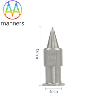
Equipment Classification Imaging-guided biopsy procedures match different needle types and imaging equipment combinations. First, ultrasound-guided conventional biopsy needles: medium-gauge standard stainless steel and polymer needles, suitable for superficial and medium-depth solid lump biopsy, with convenient operation and real-time dynamic guidance. Second, stereotactic-guided micro-biopsy needles: high-gauge ultra-thin titanium alloy needles, specially adapted for microcalcifications and tiny non-palpable lesions, realizing ultra-precise fixed-point sampling. Third, MRI-guided deep biopsy needles: long-specification reinforced needles, suitable for deep hidden lesions and complex multi-focal lesions, with strong penetration and high positioning accuracy, matching high-precision imaging guidance procedures.
Practical Operation Guide Standardized imaging-guided biopsy procedures follow unified visualized operational norms. Preoperatively, select matching imaging guidance mode according to lesion type: adopt ultrasound guidance for palpable solid lumps, stereotactic guidance for microcalcifications, and MRI guidance for deep hidden lesions. Intraoperatively, adjust patient posture to ensure clear imaging display, calibrate the puncture reference point, and plan a safe path avoiding blood vessels and nerves. Advance the biopsy needle slowly under real-time imaging monitoring, dynamically observe the needle tip position, and fine-tune the angle to ensure accurate lesion targeting. After reaching the lesion, complete multi-point sampling under imaging confirmation to ensure sample comprehensiveness. After operation, confirm no residual bleeding or abnormal tissue damage through imaging review before ending the procedure.
Practical Experience Clinical imaging-guided biopsy practice shows that visualized procedural technology increases the lesion accurate positioning rate to 100% and reduces the false-negative diagnosis rate of breast biopsy to nearly zero. It successfully solves the diagnostic dilemma of non-palpable early tiny lesions, greatly improving the early diagnosis rate of breast cancer. Real-time dynamic guidance avoids blind puncture damage, reducing intraoperative bleeding and postoperative complication rates significantly. The matching use of graded biopsy needles and imaging technology realizes accurate diagnosis of all types of breast lesions, forming a mature high-precision procedural system, which has become the mainstream standard of modern breast biopsy clinical operation.
Summary and Sublimation Imaging-guided procedural technology is a revolutionary innovation in breast biopsy operation, realizing the transformation of biopsy procedure from empirical blind operation to visualized precise operation. It makes lesion positioning, puncture path and sampling process intuitive and controllable, completely solving the accuracy and safety pain points of traditional manual biopsy procedures. The integrated system of imaging guidance and graded biopsy equipment builds a high-precision minimally invasive diagnostic platform for breast diseases, providing strong procedural support for early screening, accurate definite diagnosis and personalized treatment of breast diseases.
Future Suggestions Future imaging-guided biopsy procedures will develop towards intelligent fusion and automatic positioning. First, realize the fusion of multiple imaging technologies to improve the positioning accuracy of complex lesions. Second, develop intelligent imaging automatic path planning systems to reduce manual operation errors. Third, optimize the matching degree of biopsy needle specifications and imaging guidance modes to form exclusive procedural standards for different guidance technologies. Fourth, promote the popularization of high-precision stereotactic and MRI-guided biopsy procedures in grassroots medical institutions to improve the overall early diagnosis level of breast diseases.