How Adjustable Penetration Depth Redefines Safety Margins
Jul 24, 2026
In the rapidly evolving landscape of interventional radiology, the dogma of "one-size-fits-all" has been unequivocally debunked. The modern clinician faces a daunting array of anatomical variables-from the delicate, highly vascularized parenchyma of the thyroid to the dense, fibrous capsules of enlarged lymph nodes. It is within this context that the Adjustable penetration depth (10/20 mm) feature of our 18G Soft Tissue Biopsy Needles emerges not merely as a convenience, but as a critical evolution in patient safety and diagnostic efficacy. This functionality represents a quantum leap in engineering, addressing the nuanced demands of soft tissue sampling with unprecedented precision.
The clinical rationale for dual-depth capability is rooted in the stark variability of human pathology. Consider the thyroid gland, a butterfly-shaped organ nestled precariously close to the trachea, esophagus, and the recurrent laryngeal nerve. Nodules in this region are often minute, sometimes measuring less than a centimeter. In such scenarios, deploying a biopsy needle with a fixed 20mm cutting window is akin to performing surgery with a blindfold; the risk of transgressing the thyroid capsule and causing permanent nerve palsy or a tracheoesophageal fistula is unacceptably high. By toggling the 18G Soft Tissue Biopsy Needles to the 10mm setting, the interventional radiologist gains a mechanical "safety brake." This reduced aperture allows for the acquisition of a representative tissue core while maintaining a wide margin of safety, effectively turning a high-risk procedure into a routine outpatient intervention.
Conversely, the challenge shifts dramatically when addressing lesions in solid organs like the liver or kidneys, or when sampling fibrotic masses in the breast. Here, the 20mm setting proves indispensable. Fibrous connective tissue and neoplastic stroma are notoriously resistant to shearing forces. A shorter throw may only graze the surface of such lesions, yielding fragmented, crushed, or frankly inadequate samples that lead to inconclusive pathological reports-the dreaded "non-diagnostic" label. The 20mm depth ensures that the cutting notch fully traverses the lesion's diameter, capturing the interface between healthy and diseased tissue, which is often rich in diagnostic information for immunohistochemistry and molecular profiling.
Achieving this mechanical versatility within the compact confines of a Semi-automatic spring loaded biopsy system is a testament to sophisticated engineering. At the heart of the device lies a precision-machined cam and linkage mechanism. Unlike simpler biopsy devices that rely on a fixed-travel spring, our system incorporates a selector switch that physically alters the stop-point of the internal striker plate. When the clinician selects the 10mm option, the mechanism limits the forward travel of the inner stylet and outer cannula. This is not merely a software simulation or a visual marker; it is a hard mechanical stop that guarantees the specified depth is achieved with every trigger pull.
The true technical challenge, however, lies in maintaining consistent kinetic energy across both settings. If the spring force remains constant while the travel distance is halved, the velocity at impact would theoretically double, potentially causing excessive tissue damage or "blow-out" at the biopsy site. Conversely, if the spring is too weak for the 20mm setting, it may fail to fully deploy the cutting sheath. Our R&D team addressed this through iterative finite element analysis (FEA) and high-speed videography. We calibrated a proprietary spring alloy to deliver a specific force curve-a gradual ramp-up followed by a sustained, powerful ejection. This ensures that whether the needle travels 10mm or 20mm, the terminal velocity and shearing force remain within a narrow, clinically validated range. This consistency is verified through rigorous destructive testing, where prototypes undergo thousands of continuous firings to ensure zero deviation in sample length or morphology.
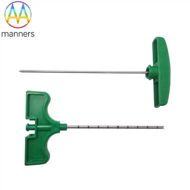
Furthermore, the integration of the optional sliding stopper elevates the safety profile even further. While the adjustable depth governs the internalmechanics of the needle, the sliding stopper provides an externalphysical barrier. In a busy interventional suite, where a physician might be managing multiple imaging screens and patient monitors simultaneously, tactile feedback is invaluable. The stopper can be locked into place at a specific point along the Centimeter markings. As the needle advances, the stopper eventually abuts against the ultrasound probe or the patient's skin, providing immediate haptic resistance. This dual-layer safety-internal mechanical limiting combined with external physical stopping-creates a redundant system that virtually eliminates the possibility of overshooting the target. For teaching hospitals and training centers, this feature is particularly valuable, allowing novice operators to develop muscle memory and spatial awareness under the protection of a mechanical safety net.
From a manufacturing perspective, producing such a device requires micron-level tolerances. The injection molding of the handle must account for thermal expansion of the plastics, ensuring the selector switch does not bind. The laser welding of the internal guide rails must be flawlessly aligned to prevent jamming during the high-G forces of firing. Each needle undergoes a final functional test where the firing mechanism is cycled multiple times to check for smooth operation. By integrating these complex mechanical principles into an ergonomic, handheld device, we provide clinicians with a tool that respects the fragility of human anatomy while delivering the robust performance required for accurate oncology. This is the essence of modern medical device design: making the complex simple, and the dangerous safe.