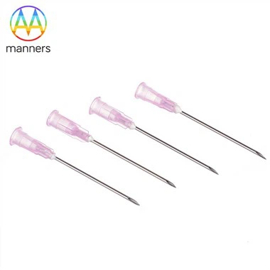
Gauge Selection
Sep 24, 2026
Pain Points
Procurement teams routinely default to "14G because everyone uses 14G," but breast biopsy is not a one-gauge-fits-all procedure. 8G, 11G, and 13G exist for large masses, vacuum-assisted biopsy, and architectural pathology where preserving glandular structures matters for treatment planning. 18G and 20G exist for superficial nodules, thin breasts, elderly patients with fragile tissue, and microcalcification clusters where cosmesis and minimal trauma are paramount. Using 14G on a 4 mm superficial lesion causes unnecessary bruising, pain, and cosmetic deformity. Conversely, using 18G on a 25 mm suspicious mass risks insufficient tissue for invasion status, forcing repeat biopsy or surgical excision. Many catalogs list gauges without clinical mapping, leaving clinicians to guess.
A second pain point is gauge inconsistency between manufacturers. One factory's 14G may measure 2.10 mm OD while another's measures 2.00 mm-a 5% difference that affects core cross-sectional area by nearly 10%. Without standardized gauge verification, "14G" becomes a moving target. A third issue is the disconnect between gauge and thin-wall design. A thin-wall 14G from a quality hypotube supplier can yield more tissue than a standard-wall 12G from a commodity mill, yet procurement compares only gauge numbers, missing the real performance differentiator. The result is hospitals overpaying for larger gauges when a well-engineered thin-wall smaller gauge would deliver equal or superior diagnostic yield.
Working Principle
Gauge is inversely proportional to diameter: lower G = bigger needle, bigger core, more trauma; higher G = smaller needle, less trauma, smaller core. Breast CNB operates mainly in 8G–20G. 8G/11G/13G are deployed in vacuum-assisted devices for large-core architectural sampling, where the thin-wall 304 or 316L tube maximizes inner diameter while maintaining pushability. 14G/15G/16G are the workhorses of standard CNB, balancing yield and cosmesis. 18G/20G serve micro-lesion and superficial applications. Thin-wall construction compresses the trauma-yield trade-off: a thin-wall 16G can approach the core volume of a standard-wall 14G with 18G-class cosmesis. Custom gauges per 2D/3D drawing or sample accommodate specialized clinical needs, such as pediatric breast cases or MRI-guided interventions with strict size constraints. The precision-ground super-sharp bevel minimizes insertion force regardless of gauge, but gauge selection still dictates the total tissue volume available for histology, immunohistochemistry, and molecular testing.
Equipment Classification
Gauge classification aligns with clinical application and manufacturing capability. 8G/11G/13G devices use large-bore thin-wall tubing, typically 316L for corrosion resistance, with vacuum-assisted or rotational cutting mechanisms. 14G/15G/16G represent the mainstream, produced on precision CNC grinders for bevel and notch, with laser etching for echogenic markings. 18G/20G require micro-drawing dies and ultra-fine grinding to maintain sharpness at small OD. Custom gauges are produced from customer 2D/3D drawings or physical samples, with full DFM (Design for Manufacturability) feedback. Manufacturing equipment includes diamond-die drawing benches for micro-OD control, CNC 5-axis grinders for bevel geometry, wire EDM for notch formation, electropolish lines for lumen finishing, and ultrasonic cleaning for bioburden reduction. Inspection uses optical comparators, projection microscopes, and tissue-simulant firing benches calibrated per gauge. All processes operate under ISO9001:2015 and ISO13485 quality systems, with SGS testing available for biocompatibility and material verification.
Practical Guide
Map lesion size, depth, breast density, and patient profile before selecting gauge. Prefer 14G/16G for most sonographic masses 8–25 mm. Use 18G for lesions under 5 mm or in very thin breasts. Use 11G/13G for vacuum-assisted architectural sampling of larger or complex lesions. Document gauge rationale in the Instructions for Use (IFU). For OEM buyers, request core-volume tables, not just gauge numbers. Validate actual OD with calibrated micrometers; reject lots outside ±0.02 mm. Consider thin-wall options to increase yield without increasing gauge. Provide 2D/3D drawings for custom gauge development with full DFM feedback. Require ISO13485 certification and lot traceability. For private-label programs, specify hub color-coding per gauge, package labeling, and sterilization method. Validate thin-wall integrity under simulated firing conditions before clinical release.
Real-World Experience
A breast program standardized 16G for lesions under 10 mm and 14G for lesions over 10 mm, replacing a previous "all 14G" policy. Insufficient-sample rate dropped from 9% to 3.4%. Patient bruising scores improved significantly despite similar diagnostic yield. In vacuum-assisted cases, 11G thin-wall devices increased core length by 18% compared to standard-wall 11G, allowing complete removal of some benign lesions during diagnostic biopsy. A separate study found that 18G micro-biopsy with echogenic guidance achieved 91% diagnostic accuracy for lesions 4–7 mm, avoiding the morbidity of larger-gauge approaches in cosmetically sensitive areas. One OEM customer switched from standard-wall 14G to thin-wall 14G 316L based on Manners' recommendation; core cross-sectional area rose 20%, and the hospital network standardized the device across 12 sites, citing improved pathologist satisfaction and reduced repeat-biopsy requests.
Summary and Elevation
Gauge is clinical grammar. Used without context, it misdiagnoses. Used with thin-wall design, echogenic marking, and clinical mapping, it personalizes breast diagnosis. The best breast biopsy programs treat gauge selection as a clinical decision, not a purchasing default. When gauge, wall thickness, notch design, bevel geometry, and echogenic marking are specified as a system, every patient receives the right needle for her specific lesion. Gauge selection is not about the number stamped on the hub; it is about matching engineering to anatomy.
Outlook and Recommendations
Lesion-sized gauge kits will replace fixed-pack procurement. Future devices will ship with 14G/16G/18G coaxial sets and AI-assisted gauge recommendation derived from ultrasound features (size, depth, density, vascularity). OEMs should develop parameterized gauge families with documented core-volume performance per thin-wall configuration. Procurement teams should evaluate vendors on clinical-outcome data, not just gauge availability and unit price. Within five years, gauge selection will be algorithm-driven, with the biopsy device automatically suggesting optimal gauge based on preoperative imaging. Manufacturers who invest in thin-wall technology, precision grinding, and clinical education will lead this transition.