From Image To Needle: Analysis Of The Core Technology Of Ultrasound-Guided Breast Biopsy
Jul 16, 2026
https://www.mayoclinic.org/tests-procedures/breast-biopsy/about/pac-20384812
The high accuracy and safety of ultrasound-guided breast biopsy lie in the integration of "visualization" and "minimally invasive" techniques. Essentially, it organically combines high-frequency ultrasound imaging technology with interventional puncture procedures to construct a complete image-guided interventional system.
First, the performance of the ultrasound imaging system is crucial. Breast-specific high-frequency linear array probes typically operate between 7.5–15 MHz, offering extremely high resolution and clearly displaying lesions down to the millimeter level. Color Doppler functionality can also identify blood flow signals within and around the lesion, helping doctors actively avoid blood vessels and reduce bleeding risk during ultrasound-guided breast biopsies. Some high-end devices have incorporated elastography and contrast-enhanced ultrasound, further improving the ability to predict the benign or malignant nature of lesions.
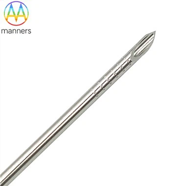
Second, the design and manufacturing process of the puncture needle are critical. The needle tip configuration directly affects the puncture feel and tissue damage: beveled needle tips (such as the Chiba needle) have strong penetrating power, suitable for rapid tissue entry; cutting needle tips (such as the Tru-Cut needle) have a "biopsy groove" with an inner core and outer sheath, allowing for the immediate removal of a complete tissue strip. In ultrasound-guided breast biopsies, the needle surface is often treated with a matte or textured finish to reduce ultrasound artifacts and improve the clarity of the needle tract imaging.
Needle specifications are equally important. Clinically, 14G–18G puncture needles are commonly used, balancing tissue acquisition volume and trauma control. Regarding length, 7 cm–15 cm is generally selected depending on the lesion depth. For deep lesions near the chest wall, longer needles are required to ensure the needle tip consistently reaches the target area.
The technical aspects of the procedure are equally important. A standard ultrasound-guided breast biopsy typically includes: preoperative ultrasound scanning for localization, marking the needle insertion point and angle, local anesthesia, needle insertion under real-time monitoring, specimen acquisition using a trigger cutting device, multiple sampling points, and postoperative compression hemostasis. Each step relies on real-time feedback from ultrasound images, especially the confirmation of the needle tip position, which must be "what you see is what you get."
In recent years, the introduction of navigation technology and automated equipment has further improved technical stability. Electromagnetic navigation systems can map the puncture needle position onto a three-dimensional reconstructed image in real time, reducing deviations caused by differences in operator experience; fully automated vacuum-assisted biopsy systems use continuous negative pressure suction to obtain multiple high-quality specimens in a single insertion, significantly improving the efficiency and diagnostic accuracy of ultrasound-guided breast biopsy. In the future, with the maturation of technologies such as AI-assisted lesion boundary identification and automatic puncture path planning, ultrasound-guided breast biopsy is expected to achieve a higher degree of intelligence and standardization, providing a solid technological foundation for the precise diagnosis and treatment of breast diseases.
Part Three: A Materials Science Perspective
The Battle at the Needle Tip: A Comprehensive Analysis of Needle Materials for Ultrasound-Guided Breast Biopsy
In ultrasound-guided breast biopsy, the puncture needle is not only an operating tool but also a core consumable that determines the quality and safety of diagnosis. The choice of different materials directly affects the strength, toughness, biocompatibility, and cost of use of the needle, making it an important direction for medical device research and development.
Stainless steel is currently the most widely used puncture needle material. 304 and 316L medical-grade stainless steel possess excellent mechanical strength and corrosion resistance, capable of withstanding repeated sterilization and high-intensity puncture operations. In ultrasound-guided breast biopsy, stainless steel needles have good rigidity and are not easily bent, making them particularly suitable for puncturing deep lesions. However, their high density and weight may increase hand fatigue for doctors during prolonged operation.
Titanium alloys (such as Ti-6Al-4V) are representative materials for high-end puncture needles. Titanium has only 60% the density of steel, making it lighter while maintaining extremely high tensile strength. More importantly, a dense oxide film naturally forms on the surface of titanium, exhibiting excellent bioinertness and significantly reducing tissue adhesion and inflammatory responses. In delicate procedures such as ultrasound-guided breast biopsy, titanium alloy needles provide more sensitive tactile feedback, improving operational accuracy. However, titanium alloys are difficult to manufacture and expensive, and are mostly used in disposable high-end products.
Medical polymer materials (such as polycarbonate (PC), polypropylene (PP), and polyetheretherketone (PEEK)) have recently emerged in the field of disposable puncture needles. These materials completely avoid the risk of metal allergies and can achieve one-piece molding of complex structures through injection molding. In ultrasound-guided breast biopsy, polymer needles are often used in conjunction with metal needle cores, with an outer cannula made of transparent or semi-transparent polymer for easy observation of tissue entry. Their biggest advantage is the complete elimination of cross-infection, and their controllable production costs make them very suitable for large-scale deployment. The choice of materials must also match the type of needle. Fine-needle aspiration (FNA) often uses stainless steel to ensure needle tip sharpness; core-needle biopsy (CNB) tends to use titanium alloys or composite materials to balance strength and weight; vacuum-assisted biopsy needles, due to their complex structure, often employ a combination design of a stainless steel body and polymer components.
Surface modification technologies are also constantly advancing. For example, titanium nitride coatings can further improve the hardness and wear resistance of stainless steel needles; hydrophilic coatings can reduce puncture resistance and minimize traction damage to surrounding tissues. These innovations are continuously improving the safety and comfort of ultrasound-guided breast biopsy.
It is foreseeable that with the development of new materials technology, puncture needles combining lightweight, high strength, high biocompatibility, and low cost will continue to emerge, bringing new possibilities to minimally invasive breast diagnosis.