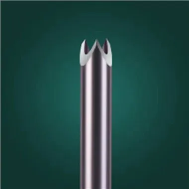
Clinical Practice - Application Skills Of Veress Needle in Laparoscopic Surgery
Jun 17, 2026
https://en.wikipedia.org/wiki/Veress_needle
The first step in laparoscopic surgery is usually to create an air leak, and the Veress needle is the key tool for accomplishing this step. The standard puncture site is located at the navel, as this is the thinnest part of the abdominal wall and there are no important blood vessels here. During the operation, the doctor first makes a skin incision about 10mm long on the lower edge of the navel using a surgical knife, then uses forceps to lift the abdominal wall and insert the Veress needle vertically or slightly towards the pelvic cavity. When the needle tip passes through the fascia and peritoneum, two distinct feelings of loss of pressure are usually felt - the first is when it breaks through the fascia, and the second is when it breaks through the peritoneum.
There are several methods to confirm that the needle tip has entered the abdominal cavity: The most common one is the "drop test" - drop a drop of normal saline at the needle tip. If negative pressure causes the water to be sucked in, it indicates that the needle tip is in the free abdominal cavity; or connect the syringe for aspiration and observe if there is blood or intestinal contents. Then connect the pneumoperitoneum machine. The initial inflation speed should be slow (1-2 liters per minute), and wait until the intra-abdominal pressure rises to 8-10 mmHg before accelerating. The entire process requires close monitoring of the pressure curve. If the pressure suddenly increases, it suggests that the needle tip may have embedded in the peritoneum or mesentery, and the position should be adjusted immediately.
In special cases, such as for patients with a history of abdominal surgery, there may be intestinal adhesions, which increases the risk of umbilical puncture. Alternative options include left subcostal puncture (Palmer point), right upper abdominal puncture, etc. In these areas, the selection of the length and angle of the Veress needle is more critical. For example, for extremely obese patients, a 150mm long needle may be needed and it should be inserted at a 45-degree angle to avoid thick subcutaneous fat.
Although intraoperative complications are rare, they should be vigilantly guarded against. The most common one is puncture injury: the incidence of intestinal perforation is approximately 0.05% - 0.2%, while vascular injury is less common but has more serious consequences. Preventive measures include: fully elevating the abdominal wall, keeping the needle tip direction away from major blood vessels, and confirming the position is correct before inflating. If injury is suspected, the inflation should be immediately stopped, and an open approach or a direct vision cannula should be used instead.
With technological advancements, some new auxiliary devices have enhanced safety. For instance, the optical Veress needle is equipped with a built-in camera, which can display the tissue layers in real time during the puncture process; ultrasound-guided punctures are also gradually becoming more widespread, especially for high-risk patients. However, no matter how advanced the tools are, solid anatomical knowledge and standardized operation procedures are always the fundamental guarantee for safety. Repeated practice on training simulators can help young doctors master the "feel" and understand the resistance changes in different tissue layers.
In conclusion, the application of the Veress needle is not only a technical task but also an art. It requires the operator to possess both courage and caution, and to make an accurate judgment within just a few seconds. Every successful puncture is a fulfillment of the promise of patient safety.