Chiba Needle In Coaxial Biopsy System
Nov 09, 2026
Pain Points of Repeated Percutaneous Biopsy
Multiple sampling is required for most suspicious lesions to raise diagnostic sensitivity, but repeated independent punctures create severe clinical pain points. Each separate puncture generates a new tissue tract, increasing cumulative bleeding risk, organ injury and tumor seeding probability. In lung biopsy, repeated transthoracic punctures sharply raise pneumothorax incidence and may require chest tube placement. Additionally, repeated skin entry points increase patient pain and procedure duration. Many operators lack a stable introducer channel; once the needle deviates, they must restart positioning and imaging scanning, wasting contrast media and CT scan time. Existing coaxial accessories are often mismatched with fine sampling needles, with poor lumen smoothness and high friction during inner needle exchange. The industry needs a compatible, low-trauma introducer needle to build a stable safe access tunnel for repeated sampling.
Working Principle of Chiba Needle as Coaxial Introducer
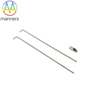
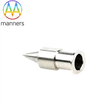
The Chiba needle serves as the outer guiding cannula in a coaxial biopsy system, leveraging its 30° bevel steerable tip and thin-wall hollow structure. The primary function is to establish a fixed percutaneous tract from skin to the margin of the target lesion. With the stylet locked inside, the Chiba needle penetrates soft tissue and parenchyma under imaging guidance. Once the tip is confirmed adjacent to the lesion, the stylet is removed, and the hollow cannula remains stationary as a stable working channel. Fine sampling needles or cutting biopsy instruments can pass through the Chiba cannula lumen to reach the lesion for multiple sampling. The bevel design allows fine adjustment of the cannula orientation during initial placement, so the whole coaxial channel aligns with the safest sampling angle, avoiding intervening vessels and airways. Medical-grade 316 stainless steel delivers smooth inner lumen surface, minimizing scratch and friction during instrument exchange.
Classification for Coaxial Application Scenarios
When used for coaxial access, Chiba needles are categorized by outer gauge matching the inner sampling device. 18G Chiba needles are the most common guiding cannula, compatible with 20G–22G fine aspiration needles and small cutting core biopsy needles. 17G Chiba cannulas are reserved for larger-bore inner instruments and thick fluid drainage. 19G Chiba needles are selected for pediatric patients or superficial high-risk lesions to lower baseline trauma. Length selection depends on lesion depth: standard 90mm for abdominal wall, liver and peripheral lung lesions, custom 120mm–150mm lengths for retroperitoneal, deep pelvic and posterior thoracic targets. All coaxial-use Chiba needles maintain the standard Chiba bevel tip; manufacturers can add depth marking lines on the cannula wall for real-time depth reading during placement. Sterile individual packaging is mandatory to preserve tract sterility during multiple instrument exchanges.
Operation Guide for Coaxial Workflow with Chiba Needle
Begin with lesion planning and safe tract design on pre-procedure CT or ultrasound images. Mark the skin entry site and administer local anesthesia. Assemble the Chiba needle with stylet, advance slowly to the lesion margin under continuous imaging verification. Confirm the cannula tip position; do not push the cannula deep into the lesion to avoid accidental vascular injury. Fix the Chiba cannula hub steadily with one hand to prevent migration, then withdraw the inner stylet. Insert the matched fine sampling needle through the Chiba cannula lumen until the tip extends out of the cannula terminal into the lesion. Perform aspiration or core sampling as required, then retract the inner sampling needle back into the cannula lumen. Repeat sampling at different lesion sub-regions while the Chiba cannula stays fixed in place. After sampling completes, withdraw the whole coaxial set and apply compression hemostasis. If tract embolization is needed for bleeding-prone patients, embolic material can be delivered through the Chiba cannula before removal.
Practical Experience in Coaxial Biopsy
The core rule is to keep the Chiba outer cannula stable after positioning; even minor hub displacement will shift the whole working channel and cause sampling failure. Operators should reserve the cannula tip at the lesion edge rather than inside the lesion, to reduce the risk of laceration when exchanging inner needles. For lung lesions, avoid advancing the cannula through bullae. Gauge matching must be strictly controlled: excessive clearance between cannula and inner needle will reduce directional control, while over-tight fit causes jamming and specimen loss. For beginners, use ultrasound first for coaxial training before CT-guided lung cases. After multiple sampling cycles, check for blood reflux inside the Chiba cannula; bright blood indicates vascular puncture and requires immediate tract management. The coaxial Chiba technique cuts puncture times drastically and is the gold standard for high-risk organ biopsy.
Conclusion & Industry Outlook
The Chiba needle's role as a coaxial introducer redefines the safety boundary of multiple-site biopsy. It solves the long-standing pain point of repeated puncture injury and stabilizes the access channel for complex interventional diagnosis. Future optimization directions include integrated depth scale, lubricant coating on the inner lumen, and color-coded hub marking for fast gauge identification. Hospital interventional suites should standardize coaxial kits pre-assembled with matched Chiba needles and sampling accessories. As precision oncology pushes multi-region molecular sampling demand, coaxial Chiba needle systems will see rising adoption in tertiary hospitals and oncology centers. Manufacturers need to strengthen compatibility testing to unify accessory interfaces and reduce intraoperative matching errors.