Applications Of Echogenic Needles in Regional Anesthesia And Nerve Blocks
Jul 05, 2026
Enhancing Precision and Safety in Brachial Plexus, Lumbar Plexus, and Fascial Plane Blocks
https://www.nature.com/articles/s41598-024-72620-8
Ultrasound-guided regional anesthesia (USGRA) is a core competency of modern anesthesiology, and needle tip visualization directly correlates with block success rates and patient safety. Traditional smooth block needles in interscalene, supraclavicular, axillary brachial plexus, and erector spinae/transversus thoracis plane blocks frequently disappear when insertion angles exceed 30°. Operators are forced to "back-infer" needle tip position from post-injection hypoechoic fluid spread, carrying risks of accidental intravascular local anesthetic injection, intraneural injection, or pneumothorax.
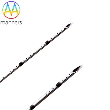
Using echogenic needles (especially 22G/21G/20G, length 50–100 mm with micro-etching or coatings), the shaft and tip display as bright white linear hyperechoic signals throughout. Anesthesiologists can observe in real time: ① whether the insertion trajectory deviates toward vessels/pleura; ② the distance from the tip to the target nerve; ③ bevel orientation for precise perineural rather than intraneural injection. Studies confirm that echogenic needles yield significantly higher tip and shaft visibility at steep insertion angles of 30°–60° compared to ordinary needles, with better operator subjective comfort and image quality scores-particularly beneficial for residents shortening their learning curves.
Clinical practice key points:
In-plane technique combined with echogenic needles delivers optimal results-full-needle visualization confirms depth; out-of-plane only shows the tip cross-section dot, but still outperforms invisibility.
Echogenic block needles often feature encrypted etched calibration rings within 20 mm of the tip, appearing as intermittent bright spots under ultrasound to assist depth estimation.
Some manufacturers offer echogenic block needles with nerve stimulation capability (electrostimulating echogenic needles), combining electrophysiological localization with ultrasound visualization for dual navigation-ideal for deep plexus blocks or obese patients.
Note: Overly strong coating echoes may produce "comet-tail artifacts" obscuring adjacent hypoechoic neural structures; gain can be appropriately lowered or etched types selected to reduce artifacts.
With the proliferation of ultrasound equipment in grassroots hospitals, equipping departments with dedicated echogenic block needles is becoming a standard quality control and teaching configuration in anesthesiology, significantly reducing nerve injury complaints and improving patient satisfaction.