Analysis Of Chiba Needle (Chiba Biopsy Needle) Production Process, Material Selection, And Medical Device Quality Certification System
Jul 04, 2026
https://radiopaedia.org/articles/chiba-needle
The Chiba needle appears simple, but its manufacturing involves precision tube processing, micro-tip grinding, electropolishing, and strict sterile quality control, which directly determine clinical puncture smoothness, tissue damage, and sampling quality.
1. Raw Material Selection
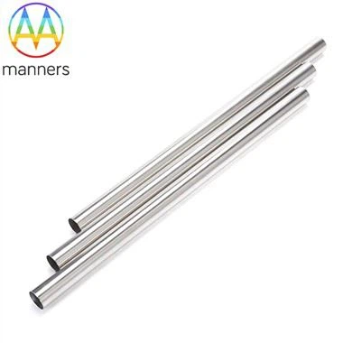
Mainstream use is medical-grade 304 or 316L stainless steel tubes (ASTM A269/A270 standards). 316L, containing molybdenum, has better corrosion resistance and good biocompatibility, suitable for short-term intra-body contact. Some high-end or MRI-compatible models use nickel-titanium (NiTi) shape memory alloy (stronger flexibility but higher cost) or non-magnetic titanium alloy. Tube outer diameter tolerance is usually controlled within ±0.02 mm, and wall thickness uniformity affects needle tip edge quality.
2. Cutting and Fixed-Length Forming
Straighten the coiled stainless steel capillary tube, then cut it to the target length (15/20/22/30 cm, etc.) using CNC cutting, and chamfer both ends to remove burrs. At this stage, tube end perpendicularity must be controlled to prevent subsequent grinding deviation.
3. Tube Body Forming and Lumen Finishing
Precision tube drawing ensures the inner lumen is smooth and unobstructed, with uniform diameter (inner diameter of 21G Chiba needle ≈0.41 mm), ensuring smooth passage of liquids/cells without retention during aspiration. Some manufacturers perform electropolishing on the inner lumen to reduce surface roughness Ra <0.2 μm.
4. Needle Tip Grinding and Bevel Forming
The core process - using a diamond wheel to grind the front end of the tube into a standard 25°–30° bevel angle, requiring a sharp edge without micro-chipping. High-end production lines use CNC five-axis grinding + online microscope inspection to ensure consistent penetration force for each needle. Some add a secondary grinding process to form an ultra-sharp edge.
5. Outer Surface Grinding and Electropolishing
The outer surface undergoes centerless grinding → electropolishing to remove machining marks, reduce insertion friction resistance, and minimize tissue drag injury. After polishing, cleaning (ultrasonic + deionized water multiple rinses) removes metal debris and polishing fluid residue.
6. Cleaning, Assembly, and Packaging
After purity testing, assemble the removable stylet or Luer connector according to product design, load into Tyvek® breathable paper-plastic pouches, sterilize with ethylene oxide (EO) or irradiation, and label with specifications (Gauge/length/batch number/expiry date).
7. Quality Control Items
Dimensional inspection: OD/ID/length/bevel angle/concentricity (projector/image measuring instrument);
Puncture force test: measure peak penetration force using simulated tissue models (e.g., silicone/animal liver);
Surface roughness: white light interferometer;
Patency: wire passing or liquid flow test;
Sterility/bacterial endotoxin/biocompatibility (cytotoxicity/sensitization/hemolysis) evaluated according to ISO 10993;
Packaging integrity: dye penetration/burst test.
8. Regulations and Certification
Regular medical Chiba needles must comply with the ISO 13485 medical device quality management system, and the product must obtain NMPA (China)/CE (EU)/FDA 510(k) (USA) registration. The packaging must be marked with warnings such as "Single-use," "Sterile," and "Do not reuse."
The clinical benefits brought by high-quality manufacturing processes are: sharp needle tip reduces pain, smooth outer surface reduces tissue damage, unobstructed inner lumen ensures cell sampling, and high batch consistency reduces operational variables - these are the fundamental reasons why interventional departments trust this instrument.