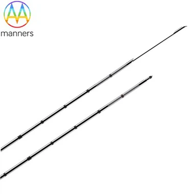
22G RF Needle For Radicular Pain
Oct 13, 2026
Pain Points
Radicular pain is one of the most common indications for lumbar RF. The patient has leg pain that follows a dermatomal pattern, and the MRI shows a disc herniation or foraminal stenosis compressing the nerve root.
The 22G RF needle is often used to treat radicular pain, but the results are variable. Some patients get excellent relief, while others get no relief at all. The variability is frustrating for the patient and the physician.
The problem is that radicular pain is not a single entity. It can be caused by mechanical compression, chemical irritation, or inflammatory mediators. Each of these causes requires a different treatment approach.
Other pain points include:
The operator may not perform a diagnostic nerve block before the RF procedure. The diagnostic block is essential for confirming that the nerve root is the source of the pain.
The operator may not use the correct RF technique. Thermal RF, pulsed RF, and dorsal root ganglion (DRG) RF are different techniques with different mechanisms of action.
The operator may not have the right needle. The needle must have the appropriate exposed tip length and insulation for the target.
Principle
Radicular pain arises from the irritation of the nerve root. The irritation can be mechanical, from compression, or chemical, from inflammatory mediators. RF energy can modulate or ablate the nerve root, reducing the pain signal.
Thermal RF creates a lesion in the nerve root, interrupting the pain signal. Pulsed RF modulates the nerve function, reducing the inflammatory response. DRG RF targets the dorsal root ganglion, which is the collection of nerve cell bodies in the dorsal root.
The physics is the same ionic friction. The RF energy heats the tissue around the tip, creating a lesion or modulating the nerve function. The key is the placement and the technique.
Device Types
Several types of 22G disposable RF needles are available for radicular pain procedures.
22G Transforaminal RF Needle is used for transforaminal RF of the nerve root. The needle is placed through the foramen, and RF energy is delivered to the nerve root.
22G Pulsed RF Root Needle is used for pulsed RF of the nerve root. The needle delivers pulsed RF energy to the nerve root, modulating the nerve function without destroying the tissue.
22G DRG RF Needle is used for DRG RF. The needle is placed in the epidural space, and the electrode is advanced to the DRG. RF energy is delivered to the DRG, interrupting the pain signal.
22G Selective Nerve Root Block Needle is used for diagnostic nerve root blocks. The needle is placed at the nerve root, and a local anesthetic is injected. The relief confirms that the nerve root is the source of the pain.
22G Combined Stimulation RF Needle is used for procedures that require sensory and motor mapping before lesioning. The needle incorporates a stimulation electrode, allowing the operator to confirm the target before delivering RF energy.
Hands-on Guide
The following steps represent best practice for using a 22G disposable RF needle for radicular pain.
Step 1: Perform a Diagnostic Nerve Root Block First. The operator should perform a diagnostic nerve root block with a local anesthetic. The patient should experience at least 50–80% relief from the block. If the patient does not get relief, RF is unlikely to be effective.
Step 2: Use Imaging to Guide the Needle. The operator should use fluoroscopy or CT to guide the needle to the target. The needle should be placed at the nerve root or the DRG.
Step 3: Perform Stimulation Testing. The operator should stimulate the nerve to confirm the target. A sensory response in the dermatome confirms that the needle is near the nerve. A motor response at a low threshold indicates that the needle is too close to a motor nerve.
Step 4: Select the Appropriate Technique. The operator should select the appropriate technique based on the clinical scenario. Thermal RF for mechanical compression, pulsed RF for inflammatory pain, and DRG RF for refractory pain.
Step 5: Deliver RF Energy. The operator should deliver RF energy at the appropriate settings for the technique. The operator should monitor the temperature and impedance throughout the procedure.
Step 6: Document the Procedure. The operator should document the diagnostic block results, the technique, the settings, and the patient's response.
Field Experience
Centers that use diagnostic nerve root blocks before RF report better patient selection and higher success rates. The diagnostic block confirms that the nerve root is the source of the pain, reducing the risk of a failed procedure.
One important lesson from the field: the diagnostic block is not always positive. Some patients with radicular pain do not get relief from the block. These patients are unlikely to respond to RF.
Another lesson: the technique matters. Thermal RF is more effective for mechanical compression, while pulsed RF is more effective for inflammatory pain. The operator should choose the technique based on the clinical scenario.
Summary
Radicular pain is a complex condition that requires a precise diagnosis and a targeted treatment. The 22G RF needle provides a minimally invasive tool for modulating or ablating the nerve root. The key is the patient selection and the technique.
The result is a procedure that is effective in the right patients and avoided in the wrong patients. The 22G needle is a precision instrument, and precision instruments require precision indications.
Outlook and Recommendations
The future of radicular pain RF will be shaped by advances in diagnosis and treatment.
Advanced Imaging. High-resolution MRI and diffusion tensor imaging will be used to identify the exact location of the nerve compression and to plan the RF procedure.
Neuromodulation Advances. Future RF techniques will combine RF with other neuromodulation modalities, such as spinal cord stimulation, to provide more comprehensive pain relief.
AI-Assisted Placement. Artificial intelligence will be used to analyze the imaging and to suggest the optimal needle trajectory.
For procurement, the recommendation is to include a diagnostic block protocol in the training bundle. The protocol should specify the local anesthetic, the volume, and the interpretation criteria.